Volume 24, Number 2—February 2018
Research Letter
Dengue-Associated Posterior Reversible Encephalopathy Syndrome, Vietnam
Figure

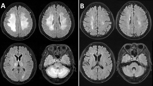
Figure. Fluid-attenuated inversion recovery magnetic resonance images of the brain of a 55-year-old woman with dengue-associated posterior reversible encephalopathy syndrome, Ho Chi Minh City, Vietnam. A) Bilateral abnormal nonenhancing, confluent high signal in the periventricular and deep cerebral white matter of the high frontal parietal area and cerebellar hemispheres, thalamus, and pons. B) Almost complete resolution of abnormal findings 7 weeks later, after treatment.
Page created: January 17, 2018
Page updated: January 17, 2018
Page reviewed: January 17, 2018
The conclusions, findings, and opinions expressed by authors contributing to this journal do not necessarily reflect the official position of the U.S. Department of Health and Human Services, the Public Health Service, the Centers for Disease Control and Prevention, or the authors' affiliated institutions. Use of trade names is for identification only and does not imply endorsement by any of the groups named above.