Volume 24, Number 4—April 2018
Synopsis
Two Infants with Presumed Congenital Zika Syndrome, Brownsville, Texas, USA, 2016–2017
Figure 1
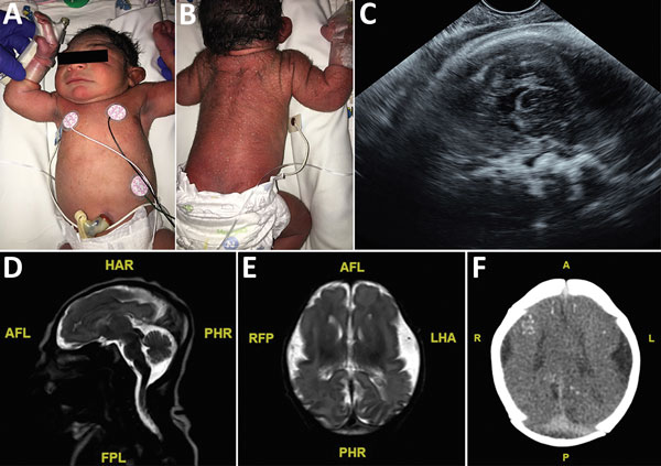
Figure 1. Term male infant (case-patient 1) with presumed congenital Zika syndrome, Brownsville, Texas, USA, 2016–2017. A) Microcephaly on the day of birth. Head circumference was 29 cm, which is 2.63 SDs below the mean value for term male newborns. Craniofacial abnormalities present are mild narrow and laterally depressed frontal bone and mild retrognathia. B) Generalized pustular melanosis rash. C) Prenatal transvaginal ultrasonographic (midsagittal plane) image at 37.2 weeks’ gestation, showing calcifications at the gray matter–white matter junction. Head circumference was 251 mm. D) Sagittal T2 magnetic resonance image on day of life 1, showing severe microcephaly, frontal lobe polymicrogyria, and hypoplastic corpus callosum. E) Axial T2 magnetic resonance image on day of life 1, showing severely hypoplastic cerebral hemispheres and corpus callosum. Symmetric frontal lobe polymicrogyria and simplified gyral pattern in the occipital and temporal lobes are present. F) Axial computed tomography image on day of life 3, showing small bilateral brain hemispheres and hypogyration of the cerebral cortex. Areas of punctate calcification located at the subcortical and gray matter–white matter junctions of the frontal, parietal, and occipital lobes are present. A, anterior; AFL, anterior left; FPL, posterior left; HAR, anterior right; L, left; LHA, left anterior; P, posterior; PHR, posterior right; R, right; RFP, right posterior.
1All authors contributed equally to this article.
2Current affiliation: Texas A&M University College of Medicine, Bryan, Texas, USA.