Volume 24, Number 6—June 2018
Dispatch
Novel Poxvirus in Proliferative Lesions of Wild Rodents in East Central Texas, USA
Figure 1
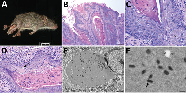
Figure 1. Histologic analysis and electron microscopy of lesions from Baiomys taylori mouse 1 infected with novel poxvirus, east central Texas, USA, 2014. A) Large epidermal masses arose from skin of both hind limbs and tail. B–D) Skin mass. Hematoxylin and eosin stain. B) The proliferative epidermis forms papillary projections with abundant hyperkeratosis and thickening of the stratum spinosum and stratum granulosum. Scale bar indicates 200 µm. C, D) Keratinocytes contain intracytoplasmic eosinophilic viral inclusions (C, arrow) that occasionally form extracellular lakes (D, arrow). Panel C scale bar indicates 20 µm; panel D scale bar indicates 50 µm. E, F) Electron microscopy. E) Cytoplasmic inclusions containing granular, electron-dense material and numerous brick-shaped virions. Scale bar indicates 1.5 µm. F) Virions have dense cores and shells of inner and outer membranes consistent with poxvirus (arrow). Scale bar indicates 300 nm.