Volume 24, Number 8—August 2018
Dispatch
Invasive Colonic Entamoebiasis in Wild Cane Toads, Australia
Figure 1
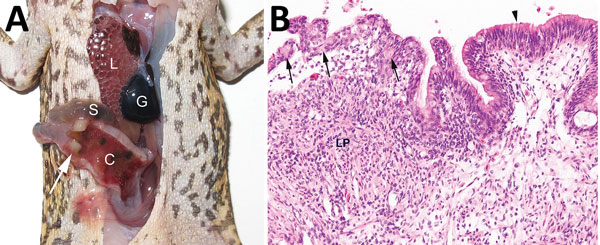
Figure 1. Invasive colonic entamoebiasis in wild cane toads (Rhinella marina), tropical Australia, 2014–2015. A) Toad with severe colonic amebiasis. The colon (C) has been opened to show intraluminal hemorrhagic content and blood clots. There is segmental full-thickness necrosis of the colon wall (white arrow). Lung (L), small intestine (S), and gall bladder (G) are annotated for perspective. B) Photomicrograph of colonic amebiasis. The affected segment of mucosal epithelium, which contains several amebae (arrows) is jumbled and sloughing from the underlying lamina propria (LP). Relatively normal colonic epithelium is present at right (arrowhead). There is lymphohistiocytic and granulocytic infiltration of the lamina propria underlying the affected epithelium. Hematoxylin and eosin stain. Original magnification ×200.