Volume 24, Number 8—August 2018
Synopsis
Abnormal Helminth Egg Development, Strange Morphology, and the Identification of Intestinal Helminth Infections
Sarah G.H. Sapp, Michael J. Yabsley, and Richard S. Bradbury
Figure 4
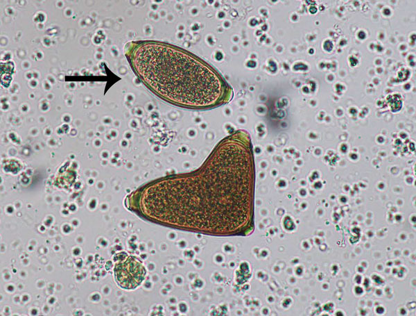
Figure 4. Conjoined Trichuris vulpis eggs from a domestic dog, visualized on fecal flotation. Arrow indicates a morphologically normal egg. Original magnification ×400. Photograph by Danielle E. Preston and courtesy of Mani Lejeune, both of the Cornell University College of Veterinary Medicine (Ithaca, NY, USA).
Page created: July 18, 2018
Page updated: July 18, 2018
Page reviewed: July 18, 2018
The conclusions, findings, and opinions expressed by authors contributing to this journal do not necessarily reflect the official position of the U.S. Department of Health and Human Services, the Public Health Service, the Centers for Disease Control and Prevention, or the authors' affiliated institutions. Use of trade names is for identification only and does not imply endorsement by any of the groups named above.