Volume 24, Number 9—September 2018
Research Letter
Transverse Myelitis and Guillain-Barré Syndrome Associated with Cat-Scratch Disease, Texas, USA, 2011
Ramia Zakhour , Pedro Mancias, Gloria Heresi, and Norma Pérez
Figure
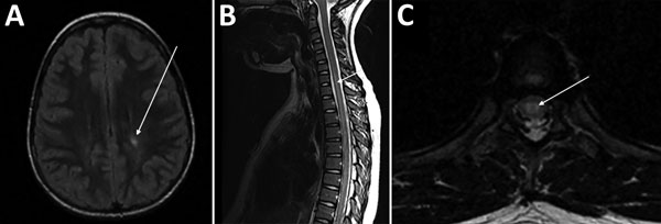
Figure. Magnetic resonance images (MRIs) on day 10 of illness in a 10-year-old girl with transverse myelitis and Guillain-Barré syndrome associated with cat-scratch disease, Houston, Texas, USA, 2011. A) Brain MRI. Arrow indicates focus of increased T2 signal in the left posterior periventricular and deep white matter. B) Sagittal spine MRI. Arrow indicates long segment of increased T2 signal centrally located within the spinal cord. C) Axial thoracic spine MRI. Arrow indicates increased central signal within the spinal cord.
Page created: August 16, 2018
Page updated: August 16, 2018
Page reviewed: August 16, 2018
The conclusions, findings, and opinions expressed by authors contributing to this journal do not necessarily reflect the official position of the U.S. Department of Health and Human Services, the Public Health Service, the Centers for Disease Control and Prevention, or the authors' affiliated institutions. Use of trade names is for identification only and does not imply endorsement by any of the groups named above.