Volume 25, Number 12—December 2019
Research Letter
Rhombencephalitis and Myeloradiculitis Caused by a European Subtype of Tick-Borne Encephalitis Virus
Lorna Neill, Anna M. Checkley, Laura A. Benjamin , M. Trent Herdman, Daniel P. Carter, Steven T. Pullan, Emma Aarons, Katie Griffiths, Bernadette Monaghan, Kushan Karunaratne, Olga Ciccarelli, Jennifer Spillane, David A.J. Moore, and Dimitri M. Kullmann
Figure
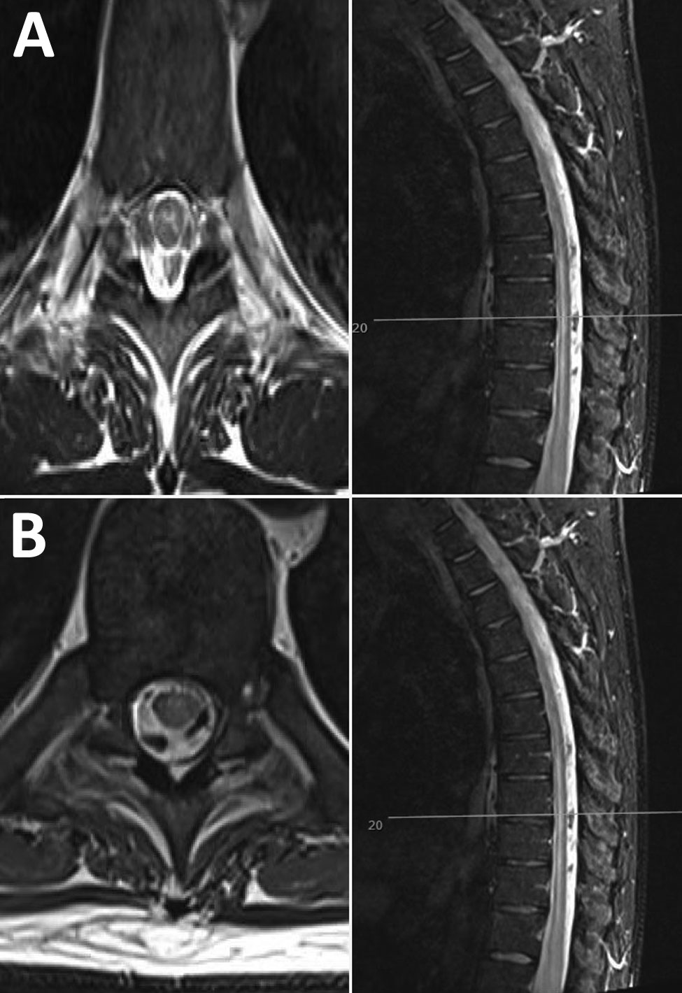
Figure. Neurologic manifestations of tick-borne encephalitis in a 38-year-old man from the United Kingdom after travel to Lithuania. A) Magnetic resonance imaging of the brain and spinal cord at onset of neurologic signs, showing possible longitudinal extensive transverse myelitis in the cervical and thoracic cord, with involvement of the central gray matter. B) One month later, increased T2 signal and mild swelling of the central gray matter of the cervical cord have both regressed, with some residual subtle signal changes throughout the spinal cord. Left, axial images; right, sagittal images.
Page created: November 18, 2019
Page updated: November 18, 2019
Page reviewed: November 18, 2019
The conclusions, findings, and opinions expressed by authors contributing to this journal do not necessarily reflect the official position of the U.S. Department of Health and Human Services, the Public Health Service, the Centers for Disease Control and Prevention, or the authors' affiliated institutions. Use of trade names is for identification only and does not imply endorsement by any of the groups named above.