Volume 25, Number 2—February 2019
Research Letter
Schistosoma haematobium–Schistosoma mansoni Hybrid Parasite in Migrant Boy, France, 2017
Figure
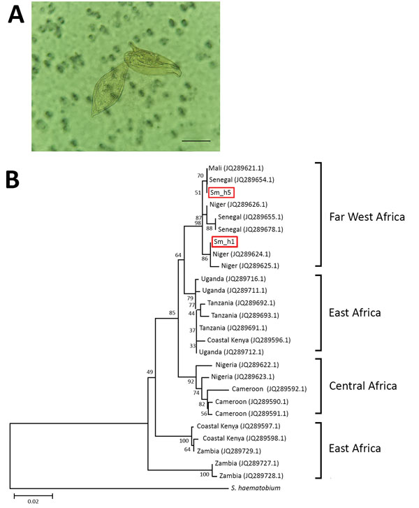
Figure. Characterization of Schistosoma parasites detected in 14-year-old migrant boy from Côte d’Ivoire in France, 2017. A) Co-detection of terminal-spined schistosome eggs (typical of Schistosoma haematobium parasites) and lateral-spined schistosome eggs (typical of Schistosoma mansoni parasites) in urine sample from migrant boy. Sample was microscopically examined after filtration. Original magnification ×400. Scale bar represents 50 µm. B) Phylogenetic analysis of S. mansoni cox1 gene haplotypes present in migrant boy (boxes). Tree was constructed by using the neighbor-joining method, the Hasegawa–Kishino–Yano plus gamma distribution model, and 1,000 bootstrap replicates. GenBank accession numbers of haplotypes sampled are provided. Scale bar indicates nucleotide substitutions per residue.