Volume 25, Number 4—April 2019
Dispatch
Pneumonic Plague in a Dog and Widespread Potential Human Exposure in a Veterinary Hospital, United States
Paula A. Schaffer1, Stephanie A. Brault1, Connor Hershkowitz1, Lauren Harris, Kristy Dowers, Jennifer House, Tawfik A. Aboellail, Paul S. Morley, and Joshua B. Daniels
Figure 1
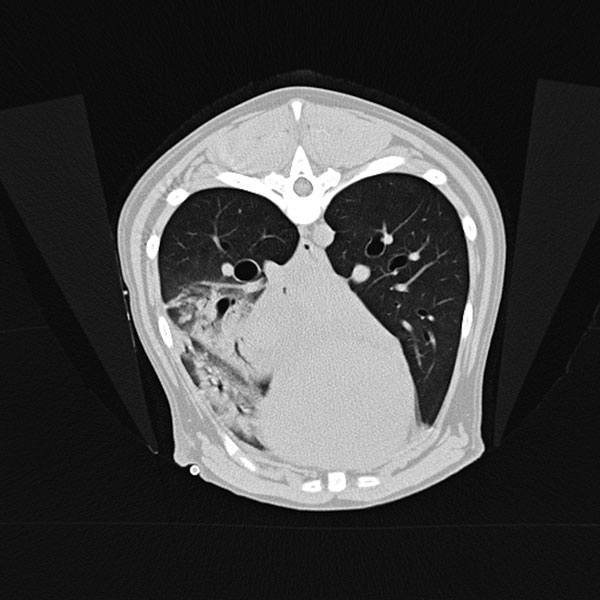
Figure 1. Transverse computed tomography of dog with pneumonic plague on day 2 of hospitalization, Colorado, USA. Image shows accessory lung lobar consolidation.
1These authors contributed equally to this article.
Page created: March 18, 2019
Page updated: March 18, 2019
Page reviewed: March 18, 2019
The conclusions, findings, and opinions expressed by authors contributing to this journal do not necessarily reflect the official position of the U.S. Department of Health and Human Services, the Public Health Service, the Centers for Disease Control and Prevention, or the authors' affiliated institutions. Use of trade names is for identification only and does not imply endorsement by any of the groups named above.