Francisella tularensis Transmission by Solid Organ Transplantation, 20171
Christina A. Nelson

, Christian Murua, Jefferson M. Jones, Kelli Mohler, Ying Zhang, Landon Wiggins, Natalie A. Kwit
2, Laurel Respicio-Kingry, Luke C. Kingry, Jeannine M. Petersen, Jennifer Brown, Saima Aslam, Melissa Krafft, Shadaba Asad, Hikmat N. Dagher, John Ham, Luis H. Medina-Garcia, Kevin Burns, Walter E. Kelley, Alison F. Hinckley, Pallavi Annambhotla, Karen Carifo, Anthony Gonzalez, Elizabeth Helsel, Joseph Iser, Michael Johnson, Curtis L. Fritz, Sridhar V. Basavaraju, and the Tularemia in Transplant Recipients Investigation Team
Author affiliations: Centers for Disease Control and Prevention, Fort Collins, Colorado, USA (C.A. Nelson, N.A. Kwit, L. Respicio-Kingry, L.C. Kingry, J.M. Petersen, A.F. Hinckley); Southern Nevada Health District, Las Vegas, Nevada, USA (C. Murua, Y. Zhang, K. Carifo, J. Iser, M. Johnson); Centers for Disease Control and Prevention, Atlanta, Georgia, USA (J.M. Jones, P. Annambhotla, S.V. Basavaraju); Phoenix Area Indian Health Service, Phoenix, Arizona, USA (K. Mohler, L. Wiggins, E. Helsel); University of California Davis Medical Center, Sacramento, California, USA (J. Brown); University of California, San Diego, California, USA (S. Aslam, M. Krafft); University Medical Center of Southern Nevada, Las Vegas (S. Asad, J. Ham, L.H. Medina-Garcia); Sunrise Hospital and Medical Center, Las Vegas (H.N. Dagher); Nevada Donor Network, Las Vegas (K. Burns); American Red Cross, Salt Lake City, Utah, USA (W.E. Kelley); University of Arizona College of Medicine, Tucson, Arizona, USA (W.E. Kelley); Sacramento County Public Health Laboratory, Sacramento (A. Gonzalez); California Department of Public Health, Sacramento (C.L. Fritz)
Main Article
Figure 2
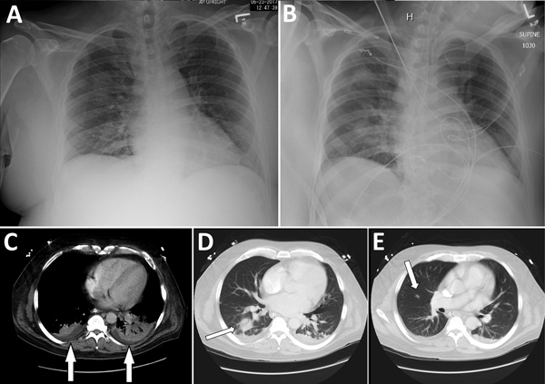
Figure 2.
Radiographs (A, B) and computed tomography (C–E) images of chest of organ donor with Francisella tularensis infection, United States, 2017. Computed tomography images were taken after brain death. A) Anteroposterior view with patient in upright position, taken on day of admission; B) anteroposterior view with patient in supine position, taken on hospital day 10. C) Small bibasilar pleural effusions with adjacent subsegmental atelectasis versus pneumonia in the lower lobes (arrows); D) 3-cm round focus of pneumonia in the right lower lobe (arrow); E) 1-cm ill-defined nodule in the inferior right upper lobe (arrow).
Main Article
Page created: March 18, 2019
Page updated: March 18, 2019
Page reviewed: March 18, 2019
The conclusions, findings, and opinions expressed by authors contributing to this journal do not necessarily reflect the official position of the U.S. Department of Health and Human Services, the Public Health Service, the Centers for Disease Control and Prevention, or the authors' affiliated institutions. Use of trade names is for identification only and does not imply endorsement by any of the groups named above.