Anthrax Epizootic in Wildlife, Bwabwata National Park, Namibia, 2017
Caitlin M. Cossaboom

, Siegfried Khaiseb, Bernard Haufiku, Puumue Katjiuanjo, Apollinaris Kannyinga, Kaiser Mbai, Thompson Shuro, Jonas Hausiku, Annety Likando, Rebekka Shikesho, Kofi Nyarko, Leigh Ann Miller, Simon Agolory, Antonio R. Vieira, Johanna S. Salzer, William A. Bower, Lindsay Campbell, Cari B. Kolton, Chung Marston, Joy Gary, Brigid C. Bollweg, Sherif R. Zaki, Alex Hoffmaster, and Henry Walke
Author affiliations: Centers for Disease Control and Prevention, Atlanta, Georgia, USA (C.M. Cossaboom, A.R. Vieira, J.S. Salzer, W.A. Bower, C.B. Kolton, C. Marston, J. Gary, B.C. Bollweg, S.R. Zaki, A. Hoffmaster, H. Walke); Republic of Namibia Ministry of Agriculture, Water, and Forestry, Windhoek, Namibia (S. Khaiseb, K. Mbai, T. Shuro); Republic of Namibia Ministry of Health and Social Services, Windhoek (B. Haufiku, P. Katjiuanjo, A. Likando, R. Shikesho, K. Nyarko); Republic of Namibia Ministry of Environment and Tourism, Windhoek (A. Kannyinga, J. Hausiku); Centers for Disease Control and Prevention, Windhoek (L.A. Miller, S. Agolory); University of Florida, Vero Beach, Florida, USA (L. Campbell)
Main Article
Figure 2
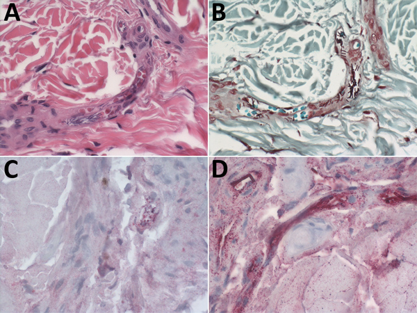
Figure 2. Photomicrographs showing hematoxylin and eosin stain and immunohistochemical findings, using assays targeting the cell wall and capsule of Bacillus anthracis, in ear-punch biopsy specimens from a hippopotamus infected with B. anthracis, Bwabwata National Park, Namibia, 2017. A) Hematoxylin and eosin stain showing large bacilli evident in vessel lumen. Original magnification × 40. B) Gram stain showing gram-variable rods evident in vessels. Original magnification × 40. C) Immunohistochemical stain of B. anthracis cell wall showing antigen evident in vessels (red). Original magnification × 40. D) Immunohistochemical stain of B. anthracis capsule showing bacilli evident in vessels (red), and bacterial antigen. Original magnification × 63.
Main Article
Page created: April 17, 2019
Page updated: April 17, 2019
Page reviewed: April 17, 2019
The conclusions, findings, and opinions expressed by authors contributing to this journal do not necessarily reflect the official position of the U.S. Department of Health and Human Services, the Public Health Service, the Centers for Disease Control and Prevention, or the authors' affiliated institutions. Use of trade names is for identification only and does not imply endorsement by any of the groups named above.