Diagnosis of Imported Monkeypox, Israel, 2018
Noam Erez
1, Hagit Achdout
1, Elad Milrot, Yuval Schwartz, Yonit Wiener-Well, Nir Paran, Boaz Politi, Hadas Tamir, Tomer Israely, Shay Weiss, Adi Beth-Din, Ohad Shifman, Ofir Israeli, Shmuel Yitzhaki, Shmuel C. Shapira, Sharon Melamed

, and Eli Schwartz
Author affiliations: Israel Institute for Biological Research, Ness-Ziona, Israel (N. Erez, H. Achdout, E. Milrot, N. Paran, B. Politi, H. Tamir, T. Israely, S. Weiss, A. Beth-Din, O. Shifman, O. Israeli, S. Yitzhaki, S.C. Shapira, S. Melamed); Shaare-Zedek Medical Center, Jerusalem, Israel (Y. Schwartz, Y. Wiener-Well); Tel Aviv University, Tel Aviv, Israel (E. Schwartz)
Main Article
Figure 2
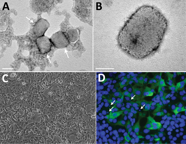
Figure 2. Transmission electron microscopy and cell culture–based diagnosis of monkeypox in patient in Israel, 2018. Virus particles were detected in lesion samples as either virion aggregates (arrows) (A) or individual virions (B) with a typical brick shape. Infected Vero cells depicted typical cytopathic effect, exhibiting cell detachment and rounding. Scale bar in A indicates 0.2 μm; scale bar in B indicates 100 nm. C) Infected Vero cells depicting typical cytopathic effect: cell detachment and rounding. Original magnification ×10. D) Immunofluorescent staining of infected Vero cells: DNA (DAPI [4′,6-diamidino-2-phenylindole] stain) and monkeypox virus; viral factories are evident (arrows). Original magnification ×25.
Main Article
Page created: April 18, 2019
Page updated: April 18, 2019
Page reviewed: April 18, 2019
The conclusions, findings, and opinions expressed by authors contributing to this journal do not necessarily reflect the official position of the U.S. Department of Health and Human Services, the Public Health Service, the Centers for Disease Control and Prevention, or the authors' affiliated institutions. Use of trade names is for identification only and does not imply endorsement by any of the groups named above.