Volume 25, Number 9—September 2019
Synopsis
Clinical Characteristics and Treatment Outcomes for Patients Infected with Mycobacterium haemophilum
Pornboonya Nookeu, Nasikarn Angkasekwinai, Suporn Foongladda, and Pakpoom Phoompoung
Figure 2
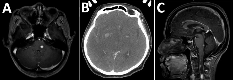
Figure 2. Imaging of brain and spine of 3 patients infected with Mycobacterium haemophilum who had involvement of the central nervous system, Bangkok, Thailand. A) Patient 1, axial T1-weighted magnetic resonance imaging scan with gadolinium showing enhanced nodule at left dorsal pons. B) Patient 2, axial contrast-enhanced computed tomography scan showing hypodensity lesions in both thalami and nodular enhancement at the bilateral basal ganglia. C) Patient 3, sagittal T1-weighted magnetic resonance imaging scan with gadolinium showing multiple enhancing nodules at dorsal pons and upper cervical cord.
Page created: August 21, 2019
Page updated: August 21, 2019
Page reviewed: August 21, 2019
The conclusions, findings, and opinions expressed by authors contributing to this journal do not necessarily reflect the official position of the U.S. Department of Health and Human Services, the Public Health Service, the Centers for Disease Control and Prevention, or the authors' affiliated institutions. Use of trade names is for identification only and does not imply endorsement by any of the groups named above.