Volume 26, Number 10—October 2020
Research Letter
Pulmonary Infection Related to Mimivirus in Patient with Primary Ciliary Dyskinesia
Cite This Article
Citation for Media
Abstract
Primary ciliary dyskinesia is a rare autosomal recessive disorder that causes oto-sino-pulmonary disease. We report a case of pulmonary infection related to mimivirus in a 10-year-old boy with primary ciliary dyskinesia that was identified using molecular techniques. Our findings indicate that the lineage C of mimivirus may cause pneumonia in humans.
In patients with primary ciliary dyskinesia (PCD), several bacterial pathogens are associated with the occurrence of pulmonary disease. However, in these patients, the many possible causative agents of pneumonia, especially viruses, have not been investigated (1). Since the detection of antibodies against mimivirus in patients with pneumonia, the potential role of mimivirus as a respiratory pathogen has been suggested (2).
In February 2017, a 10-year-old boy had severe pulmonary infection, caused by Pseudomonas aeruginosa. He had been mechanically ventilated for »10 days in the intensive care unit (ICU). One year later, he reported cough, fever, and night perspiration with excessive sputum. His physician speculated the recurrence of P. aeruginosa infection and treated him with colistin and ciprofloxacin for 3 weeks. However, symptoms of atypical pneumonia continued after 1 month.
In May 2018, care was sought agin for the child, who had excessive sputum production, weakness, chills, cough, fever, and night perspiration. He was referred to the Pasteur Institute of Iran for evaluation for nontuberculous mycobacteria. His sputum production, cough, and fever had persisted for 4 months.
The patient’s biological parameters showed elevated leukocyte count (11.9 × 1,000 cells/mL3), erythrocyte sedimentation rate (79 mm/h), and C-reactive protein (42.8 mg/L). Computed tomography scan indicated consolidation in the right lower lobe and bilateral basilar infiltrates.
Three sputum and 3 bronchoalveolar lavage (BAL) samples were sent to the laboratory for evaluation for nontuberculous mycobacteria. The results of smear, culture, and PCR for acid-fast bacilli were negative in all samples. We also evaluated what Wijers et al. found to be the most common infectious agents in PCD patients (3) but did not detect them in culture or PCR.
We used real-time PCR to identify mimivirus DNA, as previously described (4). Five (3 BAL and 2 sputum) of 6 samples were positive for mimivirus. All control samples were negative. We also sequenced the mimivirus genome using an Illumina Hiseq 2000 system (Illumina, https://www.illumina.com). Analysis of a partial genome sequence (»730 kbp) showed 99% homology to megavirus LBA111 (mimivirus lineage C) and the species Megavirus chilensis. We named the virus mimivirus PCD-1 (Appendix Figure).
Figure
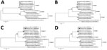
Figure. Neighbor-joining tree based on nucleotide acid sequences of mimivirus from a patient in Tehran, Iran (black circles), and reference sequences. A) The major capsid protein. B) The VV A18 helicase. C)...
To elucidate the evolutionary correlation between the mimivirus PCD-1 and other mimiviruses, we conducted phylogenetic analyses of 4 genes: the major capsid protein, the VV A18 helicase, the family B-DNA polymerase, and the D5 helicase (Figure). The phylogenetic trees indicated the close correlation of mimivirus PCD-1 with megavirus LBA111.
The role of giant viruses in human infections remains controversial. Nevertheless, small-scale reports have supported their role (2,4). We detected mimivirus DNA in sputum and BAL specimens from a 10-year-old boy with PCD in whom pneumonia developed. The negative results of culture and PCR for other pathogens strongly suggest that he had mimivirus pneumonia.
Isolation of mimivirus DNA in respiratory specimens of patients with nosocomial pneumonia who are admitted to ICUs verifies that this virus has reached the respiratory tract in these patients (5,6). The patient in this report was hospitalized in an ICU for 15 days and was mechanically ventilated for »10 days. Several studies supported the hypothesis that mimivirus occurs in pneumonia patients with ICU ventilation and is probably responsible for pneumonia and should be treated as a class 2 pathogen (2,7–9).
Although 2 previous studies have shown mimivirus infection in lower respiratory BAL specimens (2,4), we detected the virus in both upper and lower respiratory tracts, including in sputum specimens. The presence of this virus in the upper respiratory tract needs to be considered.
The first mimivirus isolated from respiratory samples belonged to lineage C (4). Consistent with this finding, the mimivirus PCD-1 from this patient was also from lineage C. In addition, another lineage C mimivirus (Shan virus) was found from the feces of a patient with pneumonia in Tunisia (10). This lineage of mimivirus may be responsible for pulmonary infection in patients; however, future research needs to confirm this result.
In summary, we detected mimivirus in a patient with primary ciliary dyskinesia who had pneumonia develop. Whether mimivirus is a causative agent of pneumonia or only extremely immunogenic is unclear, but clinicians should be aware of the potential role of this virus in human infections.
Ms. Sakhaee is a clinical microbiologist at the Pasteur Institute of Iran, Tehran, Iran. Her primary research interests focus on epidemiologic and clinical aspects of pulmonary infections.
Acknowledgment
We thank all the personnel of the Department of Mycobacteriology and Pulmonary Research, Pasteur Institute of Iran, for their assistance in this project.
References
- Alanin MC, Nielsen KG, von Buchwald C, Skov M, Aanaes K, Høiby N, et al. A longitudinal study of lung bacterial pathogens in patients with primary ciliary dyskinesia. Clin Microbiol Infect. 2015;21:1093.e1–7.
- La Scola B, Marrie TJ, Auffray J-P, Raoult D. Mimivirus in pneumonia patients. Emerg Infect Dis. 2005;11:449–52. DOIPubMedGoogle Scholar
- Wijers CD, Chmiel JF, Gaston BM. Bacterial infections in patients with primary ciliary dyskinesia: Comparison with cystic fibrosis. Chron Respir Dis. 2017;14:392–406. DOIPubMedGoogle Scholar
- Saadi H, Pagnier I, Colson P, Cherif JK, Beji M, Boughalmi M, et al. First isolation of Mimivirus in a patient with pneumonia. Clin Infect Dis. 2013;57:e127–34. DOIPubMedGoogle Scholar
- Berger P, Papazian L, Drancourt M, La Scola B, Auffray J-P, Raoult D. Ameba-associated microorganisms and diagnosis of nosocomial pneumonia. Emerg Infect Dis. 2006;12:248–55. DOIPubMedGoogle Scholar
- Bousbia S, Papazian L, Saux P, Forel J-M, Auffray J-P, Martin C, et al. Serologic prevalence of amoeba-associated microorganisms in intensive care unit pneumonia patients. PLoS One. 2013;8:
e58111 . DOIPubMedGoogle Scholar - Vincent A, La Scola B, Forel J-M, Pauly V, Raoult D, Papazian L. Clinical significance of a positive serology for mimivirus in patients presenting a suspicion of ventilator-associated pneumonia. Crit Care Med. 2009;37:111–8. DOIPubMedGoogle Scholar
- Raoult D, Renesto P, Brouqui P. Laboratory infection of a technician by mimivirus. Ann Intern Med. 2006;144:702–3. DOIPubMedGoogle Scholar
- Vanspauwen MJ, Franssen FM, Raoult D, Wouters EF, Bruggeman CA, Linssen CF. Infections with mimivirus in patients with chronic obstructive pulmonary disease. Respir Med. 2012;106:1690–4. DOIPubMedGoogle Scholar
- Saadi H, Reteno D-GI, Colson P, Aherfi S, Minodier P, Pagnier I, et al. Shan virus: a new mimivirus isolated from the stool of a Tunisian patient with pneumonia. Intervirology. 2013;56:424–9. DOIPubMedGoogle Scholar
Figure
Cite This ArticleOriginal Publication Date: September 14, 2020
Table of Contents – Volume 26, Number 10—October 2020
| EID Search Options |
|---|
|
|
|
|
|
|
Please use the form below to submit correspondence to the authors or contact them at the following address:
Abolfazl Fateh, Department of Mycobacteriology and Pulmonary Research, and Microbiology Research Center, Pasteur Institute of Iran, No. 69, 12th Farwardin Ave, Tehran, Iran
Top