Skip directly to site content Skip directly to page options Skip directly to A-Z link Skip directly to A-Z link Skip directly to A-Z link
Volume 26, Number 12—December 2020
Dispatch

Highly Pathogenic Avian Influenza A(H7N3) Virus in Poultry, United States, 2020

Sungsu Youk1, Dong-Hun Lee1Comments to Author , Mary L. Killian, Mary J. Pantin-Jackwood, David E. Swayne, and Mia K. Torchetti
Author affiliations: Southeast Poultry Research Laboratory, US Department of Agriculture Agricultural Research Service, Athens, Georgia, USA (S. Youk, M. Pantin-Jackwood, D. Swayne); University of Connecticut, Storrs, Connecticut, USA (D.-H. Lee); US Department of Agriculture, Ames, Iowa, USA (M. Killian, M. Torchetti)

Main Article

Figure 2

Median-joining phylogenetic network of the concatenated whole genome of highly pathogenic avian influenza (HPAIV) A(H7N3) viruses from South Carolina and North Carolina , USA. This network tree includes all the most parsimonious trees linking the sequences. Each unique sequence is represented by a circle sized relative to its frequency. The number of nucleotide differences between viruses is indicated on the branches. Isolates are colored according to the source premises. The black arrows indicate the H7N3 viruses with the 66-nt deletion at the neuraminidase stalk region (the NA of these 2 viruses was modified to exclude the deletion for this analysis). Both low-pathogenicity (turquoise) and HPAIV (turquoise with bolded black outline) viruses were recovered from the SC-02 premises (the hemagglutinin of the 2 HPAIVs excludes the insertion for this analysis).

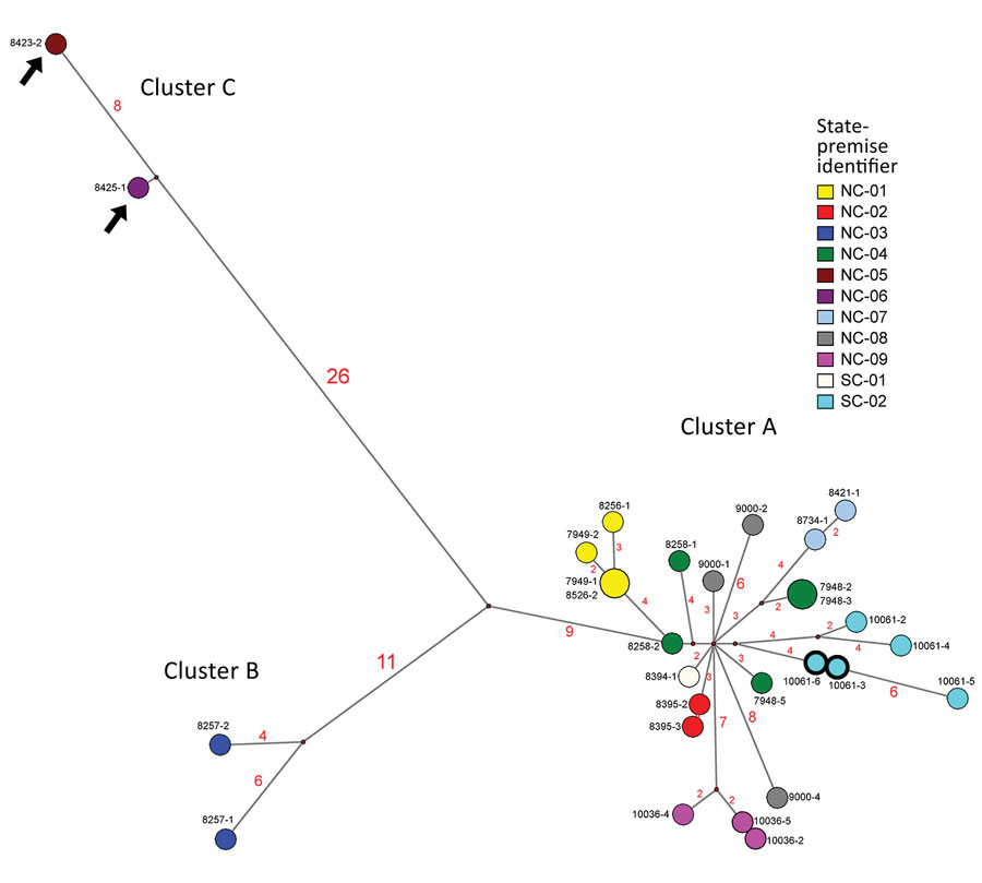
Figure 2. Median-joining phylogenetic network of the concatenated whole genome of highly pathogenic avian influenza (HPAIV) A(H7N3) viruses from South Carolina and North Carolina , USA. This network tree includes all the most parsimonious trees linking the sequences. Each unique sequence is represented by a circle sized relative to its frequency. The number of nucleotide differences between viruses is indicated on the branches. Isolates are colored according to the source premises. The black arrows indicate the H7N3 viruses with the 66-nt deletion at the neuraminidase stalk region (the NA of these 2 viruses was modified to exclude the deletion for this analysis). Both low-pathogenicity (turquoise) and HPAIV (turquoise with bolded black outline) viruses were recovered from the SC-02 premises (the hemagglutinin of the 2 HPAIVs excludes the insertion for this analysis).

Main Article

1These authors contributed equally to this article.

Page created: August 21, 2020
Page updated: November 19, 2020
Page reviewed: November 19, 2020
The conclusions, findings, and opinions expressed by authors contributing to this journal do not necessarily reflect the official position of the U.S. Department of Health and Human Services, the Public Health Service, the Centers for Disease Control and Prevention, or the authors' affiliated institutions. Use of trade names is for identification only and does not imply endorsement by any of the groups named above.
file_external