Volume 26, Number 2—February 2020
Research Letter
Hantavirus Infection with Renal Failure and Proteinuria, Colorado, USA, 2019
Swati Chand1, Sangharsha Thapa1, Shelley Kon, Steven C. Johnson, Eric M. Poeschla, Carlos Franco-Paredes, Alfonso J. Rodríguez-Morales, Salim Mattar, and Andrés F. Henao-Martínez
Figure
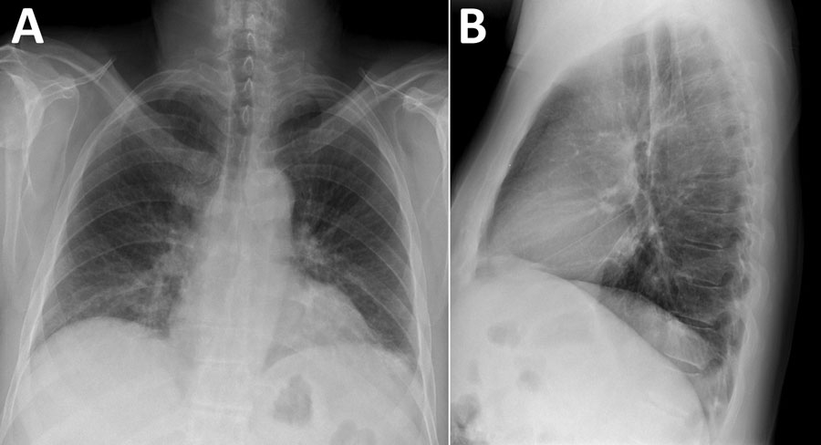
Figure. Chest radiographs displaying bibasilar patchy interstitial opacities in patient with hantavirus pulmonary syndrome, Colorado, USA. A) Posteroanterior view. B) Lateral view.
1These authors contributed equally to this article.
Page created: January 20, 2020
Page updated: January 20, 2020
Page reviewed: January 20, 2020
The conclusions, findings, and opinions expressed by authors contributing to this journal do not necessarily reflect the official position of the U.S. Department of Health and Human Services, the Public Health Service, the Centers for Disease Control and Prevention, or the authors' affiliated institutions. Use of trade names is for identification only and does not imply endorsement by any of the groups named above.