Skip directly to site content Skip directly to page options Skip directly to A-Z link Skip directly to A-Z link Skip directly to A-Z link
Volume 26, Number 6—June 2020
Dispatch

Yaws Disease Caused by Treponema pallidum subspecies pertenue in Wild Chimpanzee, Guinea, 2019

Benjamin Mubemba1, Emeline Chanove1, Kerstin Mätz-Rensing, Jan F. Gogarten, Ariane Düx, Kevin Merkel, Caroline Röthemeier, Andreas Sachse, Helene Rase, Tatyana Humle, Guillaume Banville, Marine Tchoubar, Sébastien Calvignac-Spencer, Christelle Colin, and Fabian H. LeendertzComments to Author 
Author affiliations: Copperbelt University, Kitwe, Zambia (B. Mubemba); Robert Koch Institute, Berlin, Germany (B. Mubemba, J.F. Gogarten, A. Düx, K. Merkel, C. Röthemeier, A. Sachse, S. Calvignac-Spencer, F.H. Leendertz); University of Agricultural Sciences and Veterinary Medicine, Cluj-Napoca, Romania (E. Chanove); Chimpanzee Conservation Center, Somoria, Faranah, Republic of Guinea (E. Chanove, H. Rase, G. Banville, M. Tchoubar, C. Colin); Leibniz Institute for Primate Research, Göttingen, Germany (K. Mätz-Rensing); University of Kent, Canterbury, UK (T. Humle)

Main Article

Figure 2

Histopathologic and phylogenetic analysis of yaws-like lesions and Treponema pallidum subspecies pertenue in a wild chimpanzee, Guinea. A) Histopathologic evidence suggestive of a treponemal infection. Shown here is superficial ulcerative pyogranulomatous dermatitis including formation of a mixed inflammatory cell infiltration, predominantly neutrophil granulocytes. Deeper dermal layers show the formation of a perivascular lymphocytic inflammatory cell infiltrate, focal folliculitis, and perifol

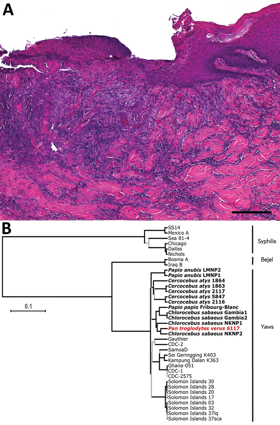
Figure 2. Histopathologic analysis of yaws-like lesions in a wild chimpanzee, Guinea, and phylogenetic placement of the Treponema pallidum subspecies pertenue strain. A) Histopathologic evidence suggestive of a treponemal infection. Shown here is superficial ulcerative pyogranulomatous dermatitis including formation of a mixed inflammatory cell infiltration, predominantly neutrophil granulocytes. Deeper dermal layers show the formation of a perivascular lymphocytic inflammatory cell infiltrate, focal folliculitis, and perifolliculitis. Skin areas adjacent to ulcerated parts show irregular epidermal hyperplasia, consistent with treponemal infections. The ulcerated areas were covered by a serocellular crust. Scale bar indicates 200 μm. B) Maximum clade credibility tree of T. pallidum strain genomes. Red indicates the chimpanzee genome generated in this study. All simian-infecting strains are shown in bold with labels showing the species of nonhuman primate, and the diseases caused by each type of bacteria are shown at right. Branches supported by posterior probabilities <0.95 in the Bayesian Markov chain Monte Carlo tree are indicated in gray. Scale bar indicates nucleotide substitutions per variable site.

Main Article

1These authors contributed equally to this article.

Page created: May 19, 2020
Page updated: May 19, 2020
Page reviewed: May 19, 2020
The conclusions, findings, and opinions expressed by authors contributing to this journal do not necessarily reflect the official position of the U.S. Department of Health and Human Services, the Public Health Service, the Centers for Disease Control and Prevention, or the authors' affiliated institutions. Use of trade names is for identification only and does not imply endorsement by any of the groups named above.
file_external