Volume 26, Number 7—July 2020
Research Letter
Urogenital Schistosomiasis in Fisherman, Nepal, 2019
Ranjit Sah , Jürg Utzinger, and Andreas Neumayr
Figure
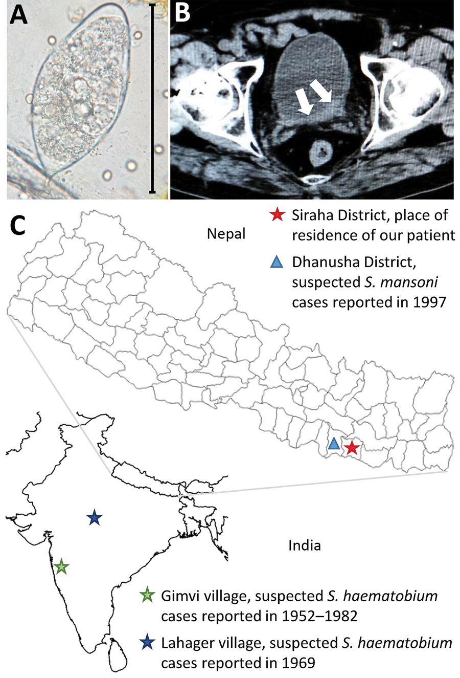
Figure. Investigation of urogenital schistosomiasis in a 34-year-old male patient in Nepal. A) Trematode egg, resembling the typical Schistosoma haematobium morphology, detected upon microscopic investigation of patient’s urine sediment. Original magnification ×40. B) Computed tomography image showing bladder wall thickening (white arrows). C) Geographic location of patient’s place of residence and of cases of human schistosomiasis reported previously in India and Nepal.
Page created: May 06, 2020
Page updated: June 18, 2020
Page reviewed: June 18, 2020
The conclusions, findings, and opinions expressed by authors contributing to this journal do not necessarily reflect the official position of the U.S. Department of Health and Human Services, the Public Health Service, the Centers for Disease Control and Prevention, or the authors' affiliated institutions. Use of trade names is for identification only and does not imply endorsement by any of the groups named above.