Volume 26, Number 7—July 2020
Research Letter
Rhabdomyolysis as Potential Late Complication Associated with COVID-19
Min Jin and Qiaoxia Tong
Figure
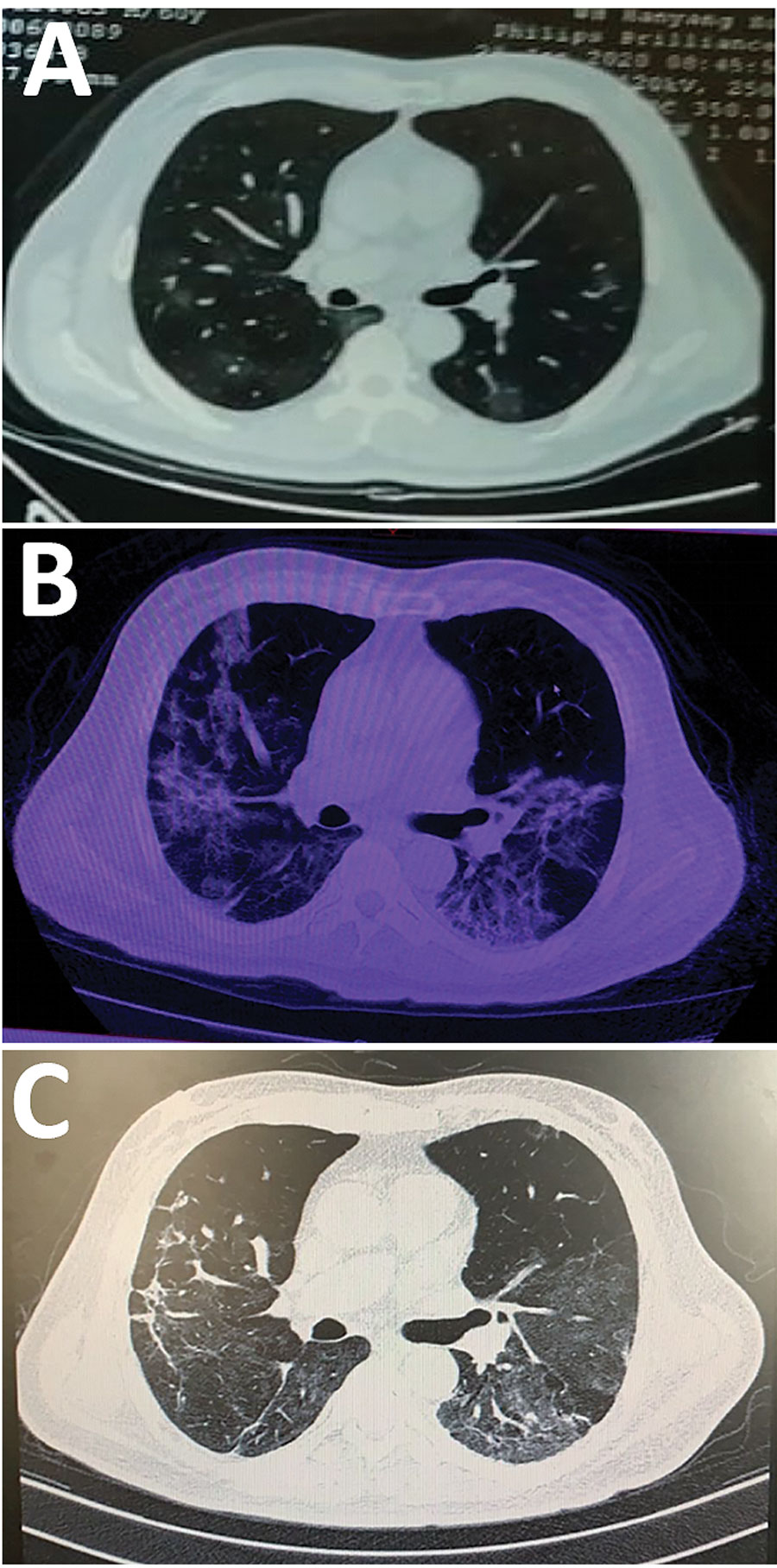
Figure. Computed tomography (CT) scan of the lungs of a 60-year-old man before and after severe acute respiratory syndrome coronavirus 2 (SARS-CoV-2) infection and rhabdomyolysis, Wuhan, China, 2020. A) CT scan before diagnosis of SARS-CoV-2 infection (3 days before hospital admission) revealed the lungs were thickened and scattered with ground-glass shadows. B) CT scan after diagnosis of SARS-CoV-2 infection with rhabdomyolysis (on hospital day 10) indicated that most of both lungs were covered with ground-glass shadows. C) CT scan after SARS-CoV-2 infection with rhabdomyolysis (on hospital day 19) indicated that pulmonary inflammation was improved.
Page created: March 20, 2020
Page updated: June 18, 2020
Page reviewed: June 18, 2020
The conclusions, findings, and opinions expressed by authors contributing to this journal do not necessarily reflect the official position of the U.S. Department of Health and Human Services, the Public Health Service, the Centers for Disease Control and Prevention, or the authors' affiliated institutions. Use of trade names is for identification only and does not imply endorsement by any of the groups named above.