Volume 26, Number 8—August 2020
Dispatch
Disseminated Echinococcus multilocularis Infection without Liver Involvement in Child, Canada, 2018
Figure 1
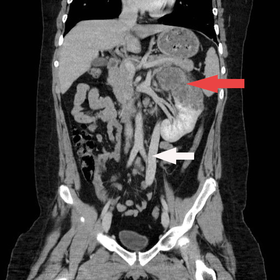
Figure 1. Coronal contrast enhanced CT (computed tomography) of the abdomen of a child with disseminated Echinococcus multilocularis infection without liver involvement, Canada, 2018. There is a large irregular hypodense left renal lesion (red arrow). A large porto-systemic shunt is partially visualized (white arrow).
1All authors contributed equally to the preparation of this article.
Page created: May 13, 2020
Page updated: July 18, 2020
Page reviewed: July 18, 2020
The conclusions, findings, and opinions expressed by authors contributing to this journal do not necessarily reflect the official position of the U.S. Department of Health and Human Services, the Public Health Service, the Centers for Disease Control and Prevention, or the authors' affiliated institutions. Use of trade names is for identification only and does not imply endorsement by any of the groups named above.