Volume 26, Number 8—August 2020
Research Letter
Cryptosporidium baileyi Pulmonary Infection in Immunocompetent Woman with Benign Neoplasm
Figure
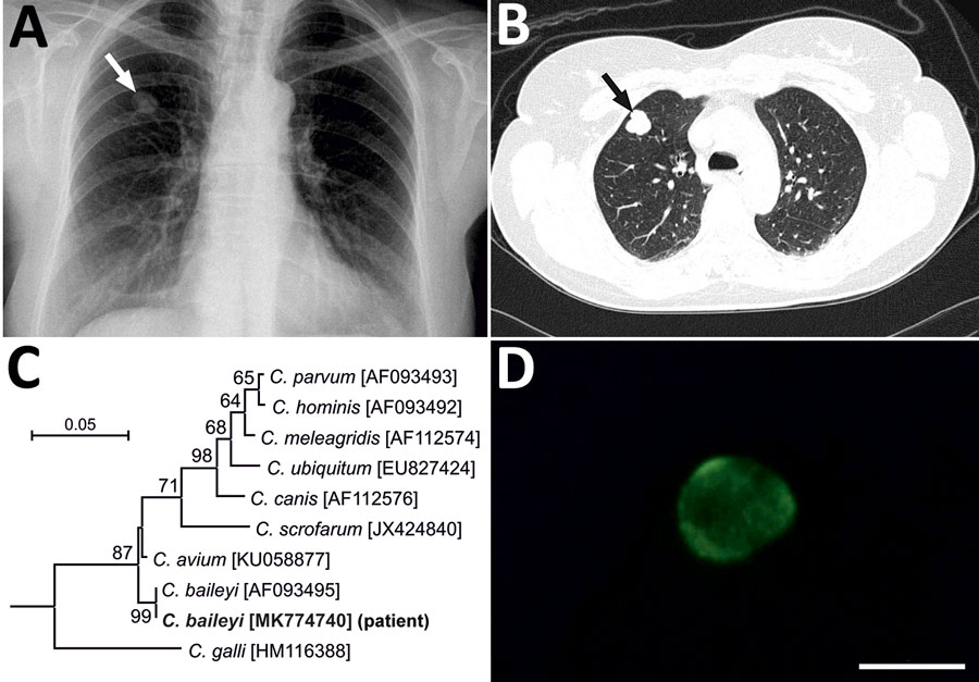
Figure. Findings from a 51-year-old immunocompetent woman with a benign neoplasm and Cryptosporidium baileyi pulmonary infection, Poland, 2015. A) Chest radiography in posterior-anterior position. A tumor, 13 × 18 mm with well-defined boundaries, is visible in the third segment of the upper right lung (arrow). B) Patient’s lung tomogram. Tumor is visible in the right lung (arrow). C) Maximum log likelihood tree based on partial sequences of gene coding small subunit rRNA of Cryptosporidium, including sequences obtained in this study (bold). Scale bar indicates nucleotide substitutions per site. D) Cryptosporidium oocyst detected in patient’s bronchial washings after immunofluorescent labeling with excitation and emission spectrum peak wave lengths of 495 nm/519 nm. Scale bar indicates 5 μm.