Skip directly to site content Skip directly to page options Skip directly to A-Z link Skip directly to A-Z link Skip directly to A-Z link
Volume 27, Number 10—October 2021
Research Letter

Emergomyces orientalis Emergomycosis Diagnosed by Metagenomic Next-Generation Sequencing

Da He1, Min Quan1, Hongyan Zhong, Zhixing Chen, Xiaohui WangComments to Author , Fang He, Junyan Qu, Taoyou Zhou, Xiaoju Lv, and Zhiyong Zong
Author affiliations: West China Hospital of Sichuan University, Chengdu, China (D. He, M. Quan, Z. Ghen, X. Wang, F. He, J. Qu, T. Zhou, X. Lv, Z. Zong); Hospital of Chengdu Office of People's Government of Tibetan Autonomous Region, Chengdu (H. Zhong); Center for Infectious Diseases, Yaan People’s Hospital, Yaan, China (X. Wang)

Main Article

Figure

Emergomyces orientalis infection in a kidney transplant patient from Tibet. A) Pulmonary consolidation with the air bronchogram sign shown on a computed tomography scan. B) Microbes stained with Grocott-Gomori's methenamine silver in the bronchoalveolar lavage fluid sample (original magnification ×1,000). C) Pathological image of 1 yeast cell shown by electron microscopy in a necrotizing granuloma from paraffin-embedded pulmonary tissue (original magnification ×16,000). D) Tiny, slightly raised white colonies on Sabouraud agar on day 20 at 25°C (left) and grayish yellow furrowed colonies on blood agar on day 30 at 35°C (right) isolated from bronchoalveolar lavage fluid samples. E) Specific secondary α-shaped conidiophore shown with fluorescent calcium staining (original magnification ×1,000). F) Ultrasound revealed a soft tissue abscess in the patient’s right subcostalis.

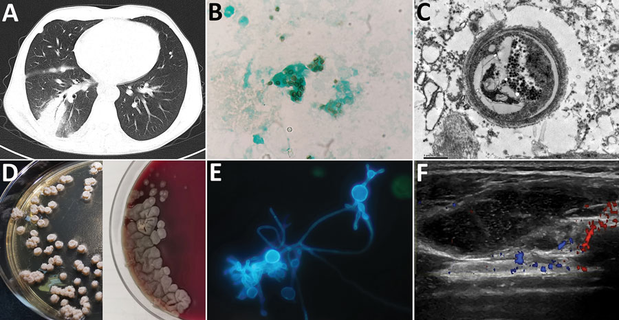
Figure. Emergomyces orientalis infection in a kidney transplant patient from Tibet. A) Pulmonary consolidation with the air bronchogram sign shown on a computed tomography scan. B) Microbes stained with Grocott-Gomori's methenamine silver in the bronchoalveolar lavage fluid sample (original magnification ×1,000). C) Pathological image of 1 yeast cell shown by electron microscopy in a necrotizing granuloma from paraffin-embedded pulmonary tissue (original magnification ×16,000). D) Tiny, slightly raised white colonies on Sabouraud agar on day 20 at 25°C (left) and grayish yellow furrowed colonies on blood agar on day 30 at 35°C (right) isolated from bronchoalveolar lavage fluid samples. E) Specific secondary α-shaped conidiophore shown with fluorescent calcium staining (original magnification ×1,000). F) Ultrasound revealed a soft tissue abscess in the patient’s right subcostalis.

Main Article

1These authors contributed equally to this article.

Page created: August 18, 2021
Page updated: October 01, 2021
Page reviewed: October 01, 2021
The conclusions, findings, and opinions expressed by authors contributing to this journal do not necessarily reflect the official position of the U.S. Department of Health and Human Services, the Public Health Service, the Centers for Disease Control and Prevention, or the authors' affiliated institutions. Use of trade names is for identification only and does not imply endorsement by any of the groups named above.
file_external