Volume 27, Number 12—December 2021
Dispatch
Evidence of Human Exposure to Tamdy Virus, Northwest China
Figure 2

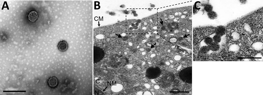
Figure 2. Visualization and subcellular localization of Tamdy virus (TAMV) virions by electron microscopy. A) Negative-staining image of purified TAMV virions. Scale bar indicates 200 nm. B) Image of Vero E6 cells infected with TAMV; arrows indicate TAMV virions in the cytoplasm. Scale bar indicates 500 nm. C) The enlarged image of interest from B. scale bar indicates 200 nm. CM, cell membrane; C, cytoplasm; NM, nuclear membrane; N, nucleus.
1These authors contributed equally to this article.
Page created: November 09, 2021
Page updated: November 19, 2021
Page reviewed: November 19, 2021
The conclusions, findings, and opinions expressed by authors contributing to this journal do not necessarily reflect the official position of the U.S. Department of Health and Human Services, the Public Health Service, the Centers for Disease Control and Prevention, or the authors' affiliated institutions. Use of trade names is for identification only and does not imply endorsement by any of the groups named above.