Volume 27, Number 3—March 2021
Research Letter
Autochthonous Case of Pulmonary Histoplasmosis, Switzerland
Yvonne Schmiedel1 , Annina E. Büchi1, Sabina Berezowska, Alexander Pöllinger, Konrad Mühlethaler, and Manuela Funke-Chambour
Figure
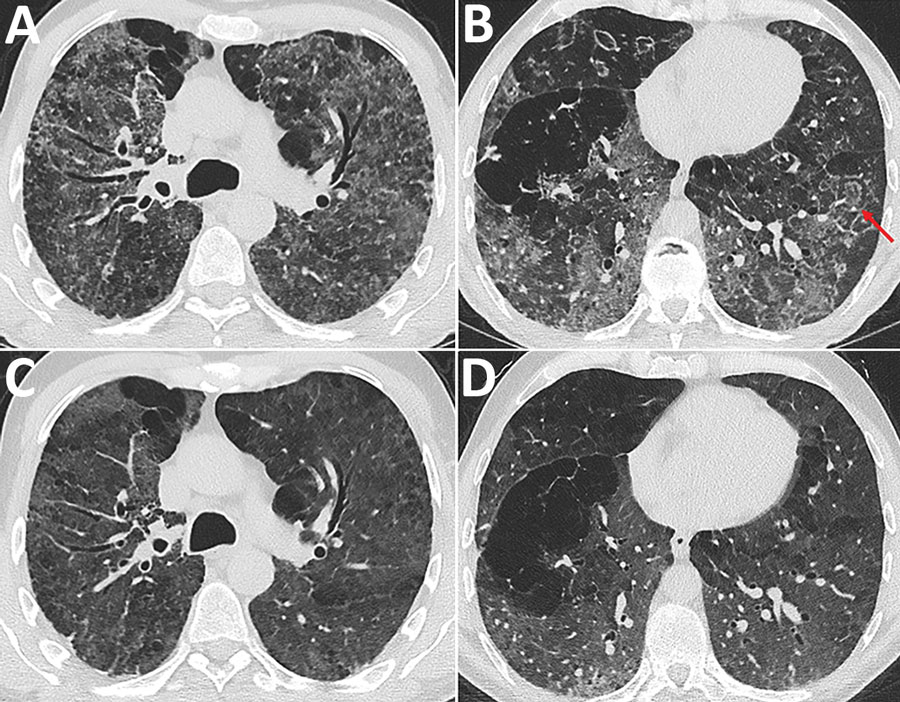
Figure. Chest computed tomography (CT) images at the level of the upper third and the lower third of the lung in a patient with pulmonary histoplasmosis, Switzerland. A, B) Initial CT shows diffuse reticulonodular pattern with ground glass opacifications, predominantly located in the upper two thirds of the lungs, and several areas with reverse halo signs (red arrows). C, D) Follow-up CT scan exhibited reduced ground-glass opacities and a regression of the micronodules. The reversed halos showed complete regression. CT, computed tomography.
1These authors contributed equally to this article.
Page created: November 10, 2020
Page updated: February 22, 2021
Page reviewed: February 22, 2021
The conclusions, findings, and opinions expressed by authors contributing to this journal do not necessarily reflect the official position of the U.S. Department of Health and Human Services, the Public Health Service, the Centers for Disease Control and Prevention, or the authors' affiliated institutions. Use of trade names is for identification only and does not imply endorsement by any of the groups named above.