Skip directly to site content Skip directly to page options Skip directly to A-Z link Skip directly to A-Z link Skip directly to A-Z link
Volume 27, Number 3—March 2021
Dispatch

Lung Pathology of Mutually Exclusive Co-infection with SARS-CoV-2 and Streptococcus pneumoniae

Tetsuya Tsukamoto1, Noriko Nakajima1, Aki Sakurai1, Masayuki Nakajima, Eiko Sakurai, Yuko Sato, Kenta Takahashi, Takayuki Kanno, Michiko Kataoka, Harutaka Katano, Mitsunaga Iwata, Yohei Doi, and Tadaki SuzukiComments to Author 
Author affiliations: Fujita Health University School of Medicine, Toyoake, Japan (T. Tsukamoto, A. Sakurai, M. Nakajima, E. Sakurai, M. Iwata, Y. Doi); National Institute of Infectious Diseases, Tokyo, Japan (N. Nakajima, Y. Sato, K. Takahashi, T. Kanno, M. Kataoka, H. Katano, T. Suzuki)

Main Article

Figure 1

Molecular detection of SARS-CoV-2 and Streptococcus pneumoniae in the lungs of a patient in Japan co-infected with both pathogens. The 42 lung sections were analyzed and the amount of SARS-CoV-2 RNA and S. pneumoniae DNA in each section was evaluated. A) The right lung was cut into 6 (R–I to R–VI); B) the left lung was cut into 7 (L–I to L–VII) coronal slices, from ventral to dorsal. Twenty-two right sections (R1–R22) in R–IV and R–V and 20 left sections (L1–L20) in L–V and L–IV are shown in black boxes. The dotted white line is the boundary between the upper and lower lobes. The SARS-CoV-2 RNA score is indicated by the number of red circles and the S. pneumoniae DNA score is indicated by the number of yellow circles. (-) indicates results under the detection limit. Scale bars indicate 2 cm. SARS-CoV-2, severe acute respiratory syndrome coronavirus 2.

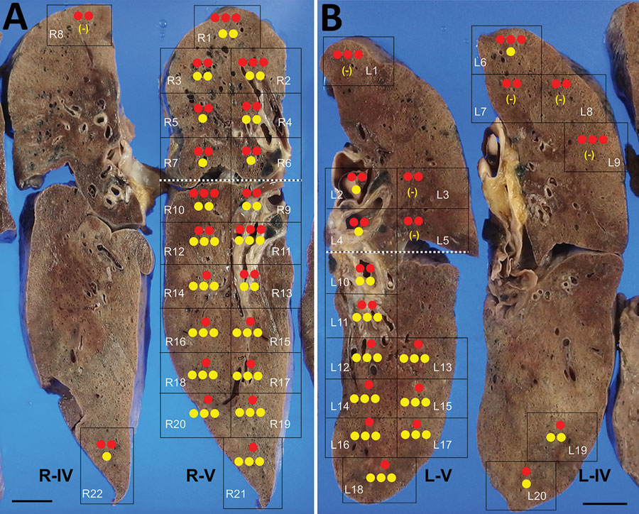
Figure 1. Molecular detection of SARS-CoV-2 and Streptococcus pneumoniae in the lungs of a patient in Japan co-infected with both pathogens. The 42 lung sections were analyzed and the amount of SARS-CoV-2 RNA and S. pneumoniae DNA in each section was evaluated. A) The right lung was cut into 6 (R–I to R–VI); B) the left lung was cut into 7 (L–I to L–VII) coronal slices, from ventral to dorsal. Twenty-two right sections (R1–R22) in R–IV and R–V and 20 left sections (L1–L20) in L–V and L–IV are shown in black boxes. The dotted white line is the boundary between the upper and lower lobes. The SARS-CoV-2 RNA score is indicated by the number of red circles and the S. pneumoniae DNA score is indicated by the number of yellow circles. (-) indicates results under the detection limit. Scale bars indicate 2 cm. SARS-CoV-2, severe acute respiratory syndrome coronavirus 2.

Main Article

1These authors contributed equally to this article.

Page created: January 06, 2021
Page updated: February 21, 2021
Page reviewed: February 21, 2021
The conclusions, findings, and opinions expressed by authors contributing to this journal do not necessarily reflect the official position of the U.S. Department of Health and Human Services, the Public Health Service, the Centers for Disease Control and Prevention, or the authors' affiliated institutions. Use of trade names is for identification only and does not imply endorsement by any of the groups named above.
file_external