Skip directly to site content Skip directly to page options Skip directly to A-Z link Skip directly to A-Z link Skip directly to A-Z link
Volume 27, Number 7—July 2021
Dispatch

Autochthonous Thelazia callipaeda Infection in Dog, New York, USA, 2020

A.B. Schwartz1Comments to Author , Manigandan Lejeune1, Guilherme G. Verocai, Rebecca Young, and Paul H. SchwartzComments to Author 
Author affiliations: Marist College, Poughkeepsie, New York, USA (A.B. Schwartz); Animal College of Veterinary Medicine at Cornell University, Ithaca, New York, USA (M. Lejeune, R. Young); Texas A&M University College of Veterinary Medicine and Biological Sciences, College Station, Texas, USA (G.G. Verocai); Center for Veterinary Care Millbrook, PC, Millbrook, New York, USA (P.H. Schwartz)

Main Article

Figure 1

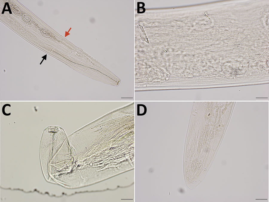
Integrated diagnostic approach for confirming Thelazia callipaeda nematodes: morphologic identification. Specimens were cleared in lactophenol before examination under an Olympus compound microscope (BX53) (https://www.olympus-lifescience.com). Images were taken with an Olympus DP73 camera, and morphometry was performed by using Olympus cellSens software. A) Cephalic end of a female worm. Black arrow indicates esophageal intestinal junction; red arrow indicates vulval opening. Original magnification ×100. B) Transverse striations (150–190/mm) in the cuticle of midbody region of a female worm. Original magnification ×200. C) Buccal cavity of a female worm, wider than deep. Note tightly spaced cuticular striations in the cephalic end. Original magnification ×200. D) Caudal end of female worm with protruding phasmids in the tip. The tail was not protruding unilaterally. Original magnification ×100.

Figure 1. Integrated diagnostic approach for confirming Thelazia callipaeda nematodes: morphologic identification. Specimens were cleared in lactophenol before examination under an Olympus compound microscope (BX53) (https://www.olympus-lifescience.com). Images were taken with an Olympus DP73 camera, and morphometry was performed by using Olympus cellSens software. A) Cephalic end of a female worm. Black arrow indicates esophageal intestinal junction; red arrow indicates vulval opening. Original magnification ×100. B) Transverse striations (150–190/mm) in the cuticle of midbody region of a female worm. Original magnification ×200. C) Buccal cavity of a female worm, wider than deep. Note tightly spaced cuticular striations in the cephalic end. Original magnification ×200. D) Caudal end of female worm with protruding phasmids in the tip. The tail was not protruding unilaterally. Original magnification ×100.

Main Article

1These authors contributed equally to this article.

Page created: May 11, 2021
Page updated: June 17, 2021
Page reviewed: June 17, 2021
The conclusions, findings, and opinions expressed by authors contributing to this journal do not necessarily reflect the official position of the U.S. Department of Health and Human Services, the Public Health Service, the Centers for Disease Control and Prevention, or the authors' affiliated institutions. Use of trade names is for identification only and does not imply endorsement by any of the groups named above.
file_external