Volume 27, Number 8—August 2021
CME ACTIVITY - Synopsis
Four Human Cases of Eastern Equine Encephalitis in Connecticut, USA, during a Larger Regional Outbreak, 2019
Figure 2
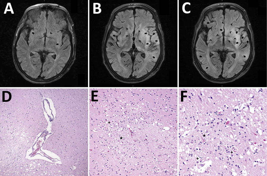
Figure 2. Mechanisms of injury in 4 human cases of Eastern equine encephalitis, Connecticut, USA, 2019. A) Magnetic resonance imaging (MRI) representative axial section from day 2 of a patient’s illness shows early development of edema around the thalamus, basal ganglia, and limbic cortical (arrows) and subcortical (arrowheads) regions. B) Representative MRI axial section from day 4 of a patient’s illness shows progression of injury in these regions and the diencephalon, basal forebrain, and subcortical areas (arrowheads). C) MRI axial section after 1 week of a patient’s illness shows expanding patchy and confluent cortical edema (arrows) and diffuse swelling in basal regions (arrowheads). D) Hematoxylin and eosin (HE)–stained photomicrograph shows the gray-white matter interface with perivascular lymphocytic cuffing and hypoxic-ischemic change in adjacent cortex. Original magnification ×40. E) HE-stained photomicrograph shows a recent gray matter microinfarction, including ischemic neurons with red cell change (5-pointed star) and perineuronal vacuolation (4-pointed star). Original magnification ×200. F) HE-stained photomicrograph shows details of acute hypoxemic-ischemic change with perineuronal (4-pointed stars) and nonspecific vacuolation, red neurons (5-pointed star), rarefaction, and pyknotic cellular debris. Original magnification ×400.
1These authors contributed equally to this article.
2Current affiliation: University of Hawai’i, Honolulu, Hawaii, USA.
3Current affiliation: University of Texas Health Science Center San Antonio, Texas, USA.