Volume 28, Number 1—January 2022
Synopsis
Outbreak of Mucormycosis in Coronavirus Disease Patients, Pune, India
Figure 2
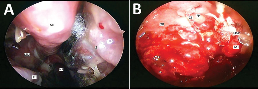
Figure 2. Diagnostic endoscopic examinations of the nasal cavities of 2 patients with mucormycosis after coronavirus disease, Pune, India. A) Right nasal cavity shows crusting in the region of the middle meatus, blackish eschar with fungal elements between the middle turbinate and the nasal septum. Necrosis has begun setting in the part of the nasal septum below the region of the eschar. Mucopurulent discharge is seen trickling from the middle meatus to the nasopharynx because of underlying sinusitis. The inferior turbinate has undergone hypertrophy because of the underlying disease. B) Left nasal cavity shows extradural abscess being drained transnasally. Multiple polyps are noted in the region of the ethmoidal fovea. The crista ethmoidalis, left middle turbinate, and the right orbital roof are destroyed because of the underlying invasion by mucor. CE, crista ethmoidalis; EDA, extradural abscess; FP, ethmoidal fovea; IT, inferior turbinate; MM, middle meatus; MT, middle turbinate; NP, nasopharynx; OR, orbital roof; S, septum.