Volume 28, Number 10—October 2022
Research Letter
Haematospirillum jordaniae Cellulitis and Bacteremia
Emil Pal, Iztok Štrumbelj, Tjaša Cerar Kišek, Marko Kolenc, Mateja Pirš, Katarina Resman Rus, Tina Triglav, and Tatjana Avšič-Županc
Figure
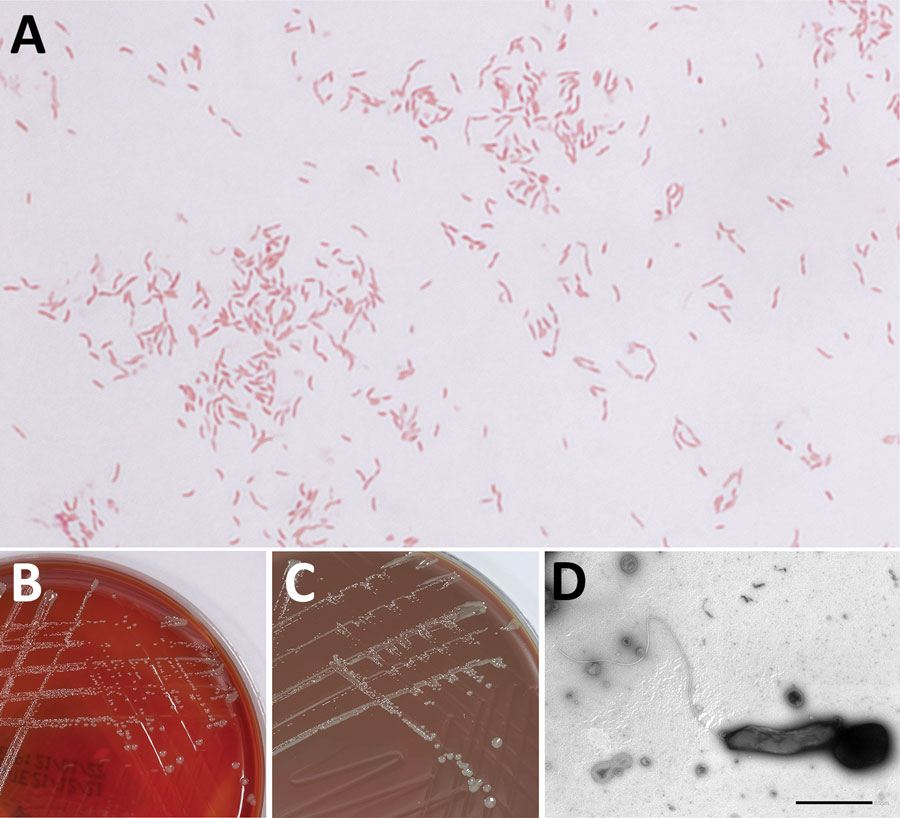
Figure. Detection of Haematospirillum jordaniae in a male patient in Slovenia. A) Gram stain of H. jordaniae; original magnification ×1,000. B) Colonies on blood agar after 3-day incubation. C) Colonies on chocolate agar after 3-day incubation. D) Transmission electron micrograph image of negatively stained cell of H. jordaniae exhibiting flagellum. Scale bar indicates 1 μm.
Page created: July 31, 2022
Page updated: September 21, 2022
Page reviewed: September 21, 2022
The conclusions, findings, and opinions expressed by authors contributing to this journal do not necessarily reflect the official position of the U.S. Department of Health and Human Services, the Public Health Service, the Centers for Disease Control and Prevention, or the authors' affiliated institutions. Use of trade names is for identification only and does not imply endorsement by any of the groups named above.