Volume 28, Number 11—November 2022
Research Letter
Serologic Evidence of Human Exposure to Ehrlichiosis Agents in Japan
Figure
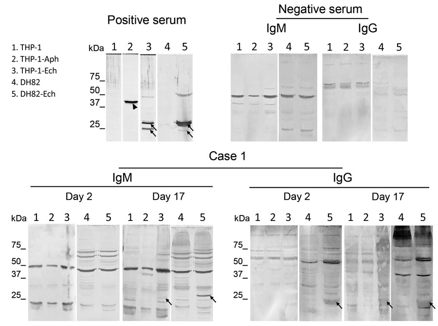
Figure. Western blots using serum samples from a febrile patient (case 1) in Wakayama Prefecture in study showing serologic evidence of human exposure to ehrlichiosis agents in Japan. Serum samples were collected from the patient on day 2 and 17 after onset of illness. Human THP-1 and canine DH82 cells were uninfected or infected with Ehrlichia chaffeensis . THP-1 cells were also infected with Anaplasma phagocytophilum. Cell lysates were separated and Western blot was performed as described (Appendix). We used uninfected THP-1 and DH82 cells as negative lysate controls. We used rabbit serum against recombinant P44 antigens specific for A. phagocytophilum and recombinant P28 antigens specific for E. chaffeensis (1:10,000 dilution) as positive serum controls. We used serum from a healthy donor as a negative control serum (Precision for Medicine, https://www.precisionbiospecimens.com). The patient’s serum samples and negative control serum were diluted 1:250 and used to probe the blots. We used alkaline-phosphatase-conjugated goat anti-human IgM μ-chain and anti-human IgG γ-chain (Thermo Fisher Scientific, https://www.thermofisher.com) as secondary antibodies. Arrows indicate E. chaffeensis-specific P28 antigens (encoded by a p28 multigene family). Arrowhead shows A. phagocytophilum-specific P44 antigen (encoded by a p44 multigene family).
1Current affiliation: Northern Fukushima Medical Center, Date, Japan.