Volume 28, Number 3—March 2022
Research Letter
Mycobacterium leprae Infection in a Wild Nine-Banded Armadillo, Nuevo León, Mexico
Lucio Vera-Cabrera , Cesar J. Ramos-Cavazos, Nathan A. Youssef, Camron M. Pearce, Carmen A. Molina-Torres, Ramiro Avalos-Ramirez, Sebastien Gagneux, Jorge Ocampo-Candiani, Mercedes Gonzalez-Juarrero, Jorge A. Mayorga-Rodriguez, Leonardo Mayorga-Garibaldi, John S. Spencer, Mary Jackson, and Charlotte Avanzi
Figure
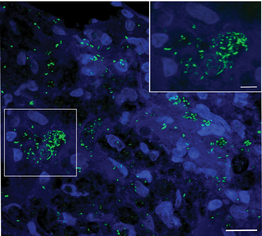
Figure. Identification and characterization of leprosy and Mycobacterium leprae acid-fast bacilli in the tissue in the wild nine-banded armadillo (Dasypus novemcinctus), Nuevo León, Mexico. SYBR gold staining shows a high density of bacilli in the spleen tissue organized in globi (boxed area at left and inset at right). Image is a merger of 16 images, 0.33 µm apart, in a z-stack taken with a 100× objective lens. Scale bars represent 20 µm (main image) and 5 µm (inset).
Page created: December 21, 2021
Page updated: February 21, 2022
Page reviewed: February 21, 2022
The conclusions, findings, and opinions expressed by authors contributing to this journal do not necessarily reflect the official position of the U.S. Department of Health and Human Services, the Public Health Service, the Centers for Disease Control and Prevention, or the authors' affiliated institutions. Use of trade names is for identification only and does not imply endorsement by any of the groups named above.