Detection of SARS-CoV-2 in Neonatal Autopsy Tissues and Placenta
Sarah Reagan-Steiner
1
, Julu Bhatnagar
1, Roosecelis B. Martines, Nicholas S. Milligan, Carly Gisondo, Frank B. Williams, Elizabeth Lee, Lindsey Estetter, Hannah Bullock, Cynthia S. Goldsmith, Pamela Fair, Julie Hand, Gillian Richardson, Kate R. Woodworth, Titilope Oduyebo, Romeo R. Galang, Rebecca Phillips, Elizaveta Belyaeva, Xiao-Ming Yin, Dana Meaney-Delman, Timothy M. Uyeki, Drucilla J. Roberts, and Sherif R. Zaki
Author affiliations: Centers for Disease Control and Prevention, Atlanta, Georgia, USA (S. Reagan-Steiner, J. Bhatnagar, R.B. Martines, E. Lee, L. Estetter, H. Bullock, C.S. Goldsmith, P. Fair, K.R. Woodworth, T. Oduyebo, R.R. Galang, D. Meaney-Delman, T.M. Uyeki, S.R. Zaki); Tulane University School of Medicine, New Orleans, Louisiana, USA (N.S. Milligan, E. Belyaeva, X.-M. Yin); Oschner Health, New Orleans (C. Gisondo, F.B. Williams, R. Phillips); Oak Ridge Institute for Science and Education, Oak Ridge, Tennessee, USA (E. Lee); Synergy America, Inc., Duluth, Georgia, USA (L. Estetter, H. Bullock); Louisiana Department of Health, Baton Rouge, Louisiana, USA (J. Hand, G. Richardson); Massachusetts General Hospital, Boston, Massachusetts, USA (D.J. Roberts)
Main Article
Figure 2

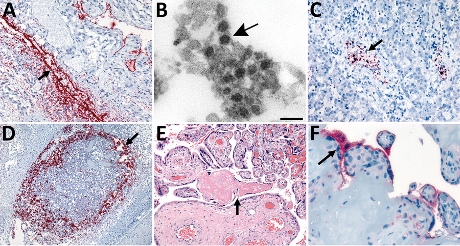
Figure 2. In situ hybridization (ISH) slides demonstrating localization of severe acute respiratory syndrome coronavirus 2 (SARS-CoV-2) genomic RNA in heart, liver, and lymph node tissues and electron microscopic evidence of viral particles in heart tissue from neonate in the United States that died with SARS-CoV-2 infection and placental histopathology and angiotensin-converting enzyme-2 immunohistochemical stain slides. A) SARS-CoV-2 RNA staining by nucleocapsid gene ISH assay in the endothelial cells in myocardium vessel walls (arrow). Original magnification ×20. B) Extracellular virus particles in the connective tissue of the heart (arrow). Scale bar indicates 100 nm. C) Intravascular staining by nucleocapsid gene ISH assay in the liver parenchyma (arrow). Original magnification ×20. D) Extensive nucleocapsid gene ISH staining within macrophages of subcapsular sinus of lymphoid follicle in the submucosa of upper airway (arrow). Original magnification ×10. E) Second trimester placenta with fibrinoid necrosis (arrow). Original magnification ×20. F) Angiotensin-converting enzyme 2 immunostaining in the membrane polarized on the maternal lake side in the syncytiotrophoblast (arrow). Original magnification ×63.
Main Article
Page created: December 02, 2021
Page updated: February 21, 2022
Page reviewed: February 21, 2022
The conclusions, findings, and opinions expressed by authors contributing to this journal do not necessarily reflect the official position of the U.S. Department of Health and Human Services, the Public Health Service, the Centers for Disease Control and Prevention, or the authors' affiliated institutions. Use of trade names is for identification only and does not imply endorsement by any of the groups named above.
