Volume 28, Number 3—March 2022
Research Letter
Subcutaneous Nodules Caused by Tropheryma whipplei Infection
Lili Wang, Peng Su, Li Song, and Lintao Sai
Figure
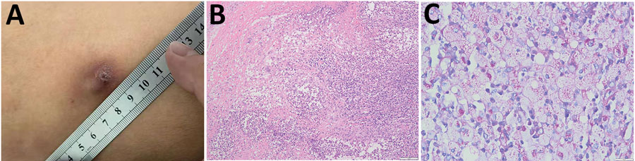
Figure. Analysis of subcutaneous nodules caused by Tropheryma whipplei infection in a patient in China, 2020. A) Surface of subcutaneous nodule on the left thigh showing ulceration. B) Histopathologic analysis showing granulomatous inflammation with massive necrosis and small abscesses formation. Periodic acid-Schiff stain. Scale bar indicates 100 μm. C) Inclusions inside cytoplasms of foamy macrophages. Periodic acid-Schiff stain. Scale bar indicates 20 μm.
Page created: January 22, 2022
Page updated: February 21, 2022
Page reviewed: February 21, 2022
The conclusions, findings, and opinions expressed by authors contributing to this journal do not necessarily reflect the official position of the U.S. Department of Health and Human Services, the Public Health Service, the Centers for Disease Control and Prevention, or the authors' affiliated institutions. Use of trade names is for identification only and does not imply endorsement by any of the groups named above.