Volume 28, Number 5—May 2022
Research Letter
Rare Case of Rickettsiosis Caused by Rickettsia monacensis, Portugal, 2021
Cite This Article
Citation for Media
Abstract
We report a case of rickettsiosis caused by Rickettsia monacensis in an immunocompetent 67-year-old man in Portugal who had eschar, erythematous rash, and an attached Ixodes ricinus tick. Seroconversion and eschar biopsy led to confirmed diagnosis by PCR. Physicians should be aware of this rare rickettsiosis, especially in geographic regions with the vector.
Rickettsia monacensis, spotted fever group rickettsiae (SFGR), are bacteria transmitted by Ixodes spp. ticks and are rarely reported as causing disease in humans. Few cases have been documented and laboratory confirmed (1–4). R. monacensis infection causing Mediterranean spotted fever (MSF)–like rickettsiosis was described in 2007 for 2 patients from La Rioja and the Basque Country, Spain, followed by 1 case in Italy (2012) and 2 cases in South Korea (2017 and 2019) (1–4). Despite the few human infections described, R. monacensis is frequently found (0.5%– 42.5%) in Ixodes ricinus ticks in Europe, including Portugal and North Africa, and in another Ixodes species tick in Asia (3–5).
Three previously reported rickettsioses in Portugal were MSF caused by R. conorii, tickborne lymphadenopathy caused by R. slovaca, and lymphangitis-associated rickettsiosis caused by R. sibirica mongolitimonae (6–8). We report R. monacensis infection in a human and Rickettsia in the attached tick.
Figure
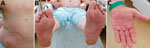
Figure. Patient with rickettsiosis caused by Rickettsia monacensis, Portugal, 2021. A) Rash and eschar; B) rash on soles; C) rash on palms.
In February 2021, a 67-year-old man with alcoholism–associated dilated cardiomyopathy and diabetes mellitus type 2 was hospitalized in Lisbon, Portugal. The patient reported a 5-day history of fever and appearance of rash on day 3 of fever onset. He lived in Lisbon and had traveled to a rural area 5 days before symptom onset. At admission, he had fever, fatigue, myalgia, and anorexia. Physical examination showed disperse upper-body erythematous exanthema, palmo-plantar erythema, and an eschar surrounded by erythema on his upper left back (Figure). An engorged female I. ricinus tick was removed from the patient. Laboratory evaluation showed hematologic, hepatic, and renal abnormalities; anemia (hemoglobin 9.7 g/dL); lymphopenia (420 cells/μL); thrombocytopenia (38,000 platelets/mm3); and increased serum levels of creatinine (2.23 mg/dL), alanine aminotransferase (73 IU/L), aspartate aminotransferase (89 IU/L), creatine phosphokinase (116 IU/dL), lactate dehydrogenase (148 IU/L), and C-reactive protein (159.5 mg/L). Electrocardiography findings were unremarkable. Oral doxycycline (200 mg/d) was empirically started on hospitalization day 1.
After the patient had been hospitalized for 12 hours and received 1 dose of doxycycline, we biopsied the eschar and collected a blood sample. PCR and DNA sequence analysis of partial fragments of ompA and gltA genes from the tick and biopsy samples showed 100% identity with nucleotide sequences of R. monacensis (GenBank accession no. LN794217). Screening for Borrelia DNA in the tick was negative.
For antibody testing we used an immunofluorescence assay from FOCUS Diagnostics (https://www.focusdx.com), which used commercial R. conorii IFA substrate slides for IgG and IgM; results demonstrated seroconversion within 2 weeks in consecutively collected samples. We detected no antibodies in the acute-phase serum sample collected on day 6 after symptom onset, and we detected reactive antibodies against SFGR (IgM titer 32, IgG titer 128) in the second sample only, collected 3 weeks after illness onset (9). Supplemental methods and results are in the Appendix.
After 48 hours of antimicrobial therapy, the patient was afebrile; after 4 days, exanthema was completely resolved; and after 7 days, all symptoms had resolved. The patient was discharged and scheduled for outpatient follow-up.
We confirm that R. monacensis caused disease in this patient. Very few cases of human infection with R. monacensis have been reported, possibly because this species is not considered to be very pathogenic and for most patients might cause self-limited infection (1–5). Another hypothesis is that cases have been misdiagnosed or confirmed by serology only, which cannot distinguish among SFGR species (8,9). Moreover, if cases occur in the autumn/winter, when adult I. ricinus ticks are more active and outside the peak season (June–September) for MSF, some physicians might not think of rickettsiosis as the cause, particularly if there is no epidemiologic context and clinical findings are not highly suggestive.
For the patient reported here, we identified an eschar, as was done for the 3 other patients from Italy and South Korea (Table). However, the first 2 patients identified in Spain did not have any sign of an eschar. We are unaware whether any specific patient host factors could be associated with R. monacensis infection, but alcoholism in the patient reported here could have been a risk factor for severity (8). With exception of the patient from Italy, all patients were >59 years of age, including the patient from Portugal, and at least 3 were hospitalized. In general, it would seem that older persons are more susceptible to disease, even when infected with low-pathogenicity Rickettsia. For instance, in the case report of an 8-year-old child from Croatia with Lyme borreliosis, in whom R. monacensis DNA was also detected in a skin biopsy of the erythema migrans tissue, antibodies against Borrelia were detected but not antibodies against SFGR (10).
This case of infection with R. monacensis, formerly considered to be of low pathogenicity and found in Ixodes spp. ticks, was associated with disease in an immunocompetent patient. Other cases may be underdiagnosed, particularly outside the usual summer months when MSF cases peak in Portugal. Moreover, because R. monacensis shares the same vector as Borrelia spp. and these co-infections have been detected, physicians should be aware of this rickettsiosis, especially in areas where the vector is present.
Dr. de Sousa is a public health microbiologist responsible for the Rickettsial and the Gastrointestinal Viral Infections Units at the National Institute of Health Dr. Ricardo Jorge. She works on research and diagnosis of rickettsial diseases.
References
- Jado I, Oteo JA, Aldámiz M, Gil H, Escudero R, Ibarra V, et al. Rickettsia monacensis and human disease, Spain. Emerg Infect Dis. 2007;13:1405–7. DOIPubMedGoogle Scholar
- Madeddu G, Mancini F, Caddeo A, Ciervo A, Babudieri S, Maida I, et al. Rickettsia monacensis as cause of Mediterranean spotted fever-like illness, Italy. Emerg Infect Dis. 2012;18:702–4. DOIPubMedGoogle Scholar
- Kim YS, Choi YJ, Lee KM, Ahn KJ, Kim HC, Klein T, et al. First isolation of Rickettsia monacensis from a patient in South Korea. Microbiol Immunol. 2017;61:258–63. DOIPubMedGoogle Scholar
- Kim SW, Kim CM, Kim DM, Yun NR. Case report: coinfection with Rickettsia monacensis and Oraientia tsutsugamushi. Am J Trop Med Hyg. 2019;101:332–5. DOIPubMedGoogle Scholar
- Parola P, Paddock CD, Socolovschi C, Labruna MB, Mediannikov O, Kernif T, et al. Update on tick-borne rickettsioses around the world: a geographic approach. Clin Microbiol Rev. 2013;26:657–702. DOIPubMedGoogle Scholar
- de Sousa R, Pereira BI, Nazareth C, Cabral S, Ventura C, Crespo P, et al. Rickettsia slovaca infection in humans, Portugal. Emerg Infect Dis. 2013;19:1627–9. DOIPubMedGoogle Scholar
- de Sousa R, Barata C, Vitorino L, Santos-Silva M, Carrapato C, Torgal J, et al. Rickettsia sibirica isolation from a patient and detection in ticks, Portugal. Emerg Infect Dis. 2006;12:1103–8. DOIPubMedGoogle Scholar
- Sousa R, França A, Dória Nòbrega S, Belo A, Amaro M, Abreu T, et al. Host- and microbe-related risk factors for and pathophysiology of fatal Rickettsia conorii infection in Portuguese patients. J Infect Dis. 2008;198:576–85. DOIPubMedGoogle Scholar
- Portillo A, de Sousa R, Santibáñez S, Duarte A, Edouard S, Fonseca IP, et al. Guidelines for the detection of Rickettsia spp. Vector Borne Zoonotic Dis. 2017;17:23–32. DOIPubMedGoogle Scholar
- Tijsse-Klasen E, Sprong H, Pandak N. Co-infection of Borrelia burgdorferi sensu lato and Rickettsia species in ticks and in an erythema migrans patient. Parasit Vectors. 2013;6:347. DOIPubMedGoogle Scholar
Figure
Table
Cite This ArticleOriginal Publication Date: April 11, 2022
Table of Contents – Volume 28, Number 5—May 2022
| EID Search Options |
|---|
|
|
|
|
|
|
Please use the form below to submit correspondence to the authors or contact them at the following address:
Rita de Sousa, Av. Da Liberdade n°5, 2965, Águas de Moura, Portugal
Top