Volume 28, Number 5—May 2022
Research Letter
Angiostrongylus cantonensis in a Red Ruffed Lemur at a Zoo, Louisiana, USA
Jessica Rizor, Ryan A. Yanez, Tuddow Thaiwong, and Matti Kiupel
Figure
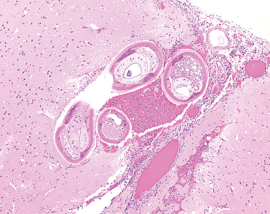
Figure. Formalin-fixed brainstem specimen from red ruffed lemur (Varecia rubra) infected with Angiostrongylus cantonensis nematodes at a zoo in Louisiana, USA. Hematoxylin and eosin stain shows adult nematodes measuring ≈50–70 μm in diameter with 3–4 μm thick, smooth, eosinophilic cuticle and prominent lateral cords. Nematodes have a coelomyarain musculature and a pseudocoelom that contains a reproductive tract and an intestinal tract, lined by multinucleated cells. Original magnification ×10.
Page created: March 11, 2022
Page updated: April 19, 2022
Page reviewed: April 19, 2022
The conclusions, findings, and opinions expressed by authors contributing to this journal do not necessarily reflect the official position of the U.S. Department of Health and Human Services, the Public Health Service, the Centers for Disease Control and Prevention, or the authors' affiliated institutions. Use of trade names is for identification only and does not imply endorsement by any of the groups named above.