Volume 28, Number 6—June 2022
Research Letter
Molecular Diagnosis of Pseudoterranova decipiens Sensu Stricto Infections, South Korea, 2002‒2020
Cite This Article
Citation for Media
Abstract
Human Pseudoterranova decipiens larval infections were diagnosed by molecular analysis of mitochondrial cox1 and nd1 genes in 12 health check-up patients in South Korea during 2002–2020. Based on high genetic identity (99.3%–100% for cox1 and 96.7%–98.0% for nd1), we identified all 12 larvae as P. decipiens sensu stricto.
Human anisakiasis, which is caused by infection with larvae of the family Anisakidae after consuming infested marine fish or squids, is one of the most serious foodborne zoonotic diseases (1). Several species of Anisakis (A. simplex sensu stricto, A. physeteris, and A. pegreffii) (1–3), Pseudoterranova (P. decipiens sensu stricto, P. azarasi, and P. cattani) (4–6), and Contracecum (C. osculatum) (7) nematodes have been reported to cause human infections.
Human anisakiasis was reported in the Netherlands during 1960 and has been found to occur in various parts of the world, including Japan and South Korea (1). Most human case-patients were infected with larvae of A. simplex s.s. (1). However, after 1999, a considerable number of cases infected with A. pegreffii nematodes (a sibling species of A. simplex s.s.) were diagnosed in Italy, Japan, and South Korea on the basis of molecular analysis of the larvae (2,3). Compared with Anisakis spp. nematodes, human infections with Pseudoterranova spp. nematodes have been relatively rare in Asia (1,4–6). In South Korea, among 645 anisakidosis cases recorded after 1971 until 2015, only ≈11.8% were infected with Pseudoterranova larvae (8). However, all of these Pseudoterranova infections were diagnosed on the basis of only the morphology of the larvae (8).
Within the genus Pseudoterranova, 8 species have been validated on the basis of molecular and morphologic/biologic characteristics: P. decipiens s.s., P. kogiae, P. ceticola, P. azarasi, P. krabbei, P. bulbosa, P. decipiens E, and P. cattani (9). Among those, 6 species (P. decipiens s.s., P. krabbei, P. bulbosa, P. azarasi, P. decipiens E, and P. cattani) are morphologically and biologically related to each another and designated as the P. decipiens species complex or P. decipiens sensu lato (9). These species can be discriminated by allozyme or molecular genetic analyses (10).
In our study, 12 human pseudoterranoviasis cases were found among patients who visited health check-up centers or hospitals in South Korea during 2002–2020 because of vague abdominal discomfort. Larvae were extracted by using gastrointestinal endoscopy (11 case-patients) or colonoscopy (1 case-patient). The larvae were confirmed to be P. decipiens s.s. by sequence analysis of the mitochondrial cytochrome c oxidase 1 (cox1) and NADH dehydrogenase subunit 1 (nd1) genes.
The patients consisted of 5 men (41–55 years of age) and 7 women (29–59 years of age). A total of 12 larvae (1 larva from each patient) were collected from the stomach (11 patients) or cecum (1 patient) (Appendix Table 1) and were processed for sequencing of 2 mitochondrial genes (Appendix).
Sequences of the cox1 (141 bp) (samples nos. OK539788–OK539799) and nd1 (153 bp) genes (OK539800–OK539807) showed high homologies with the sequences of P. decipiens s.s. (GenBank accession no. NC_031645 for cox1 and nd1). The homology between samples from this study and P. decipiens s.s. was 99.3%–100% for cox1 and 97.4%–98.0% for nd1 (Appendix Tables 1–3).
Figure
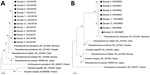
Figure. Phylogenetic analyses of Pseudoterranovanematode larvae extracted from 12 health check-up patients in South Korea, 2002–2020 (black dots), in comparison with other anisakid species. A) mitochondrial cytochrome oxidase c...
The phylogenetic tree for cox1 showed that the 12 study samples were tightly clustered with P. decipiens s.s. reported from Germany but separate from P. bulbosa from Canada, P. cattani from Chile, P. krabbei from Norway, and P. azarasi from Japan (Figure). The phylogenetic tree for nd1 showed that 8 study samples were closely aligned with P. decipiens s.s. reported from Germany but clearly separated from P. cattani from Chile, P. bulbosa from Canada, P. krabbei from Norway, and P. azarasi from Japan (Figure). We also determined genetic distances between the study specimens and P. decipiens, P. azarasi, P. bulbosa, P. cattani, and P. krabbei for cox1 (Appendix Table 2) and nd1 (Appendix Table 3).
For the specific diagnosis of anisakid larvae, analysis of the larval morphology is highly useful. However, extracting a fully intact larva from human patients for high-quality morphologic analysis is usually difficult. In such instances, molecular analysis of the larvae is helpful and essential for obtaining a specific diagnosis. Analyses of the internal transcribed spacer region and partial 28S rDNA could discriminate P. decipiens s.s. from P. bulbosa, P. krabbei, P. cattani, and possibly P. decipiens E (10). However, great sequence similarity was observed between P. decipiens s.s. and P. azarasi. Thus, it was difficult to distinguish them by using nuclear genes (10). Some investigators used mitochondrial genes, including cox1, cox2, and nd1, to distinguish them (4,5).
In our study, we used 2 mitochondrial genes, cox1 and nd1, to distinguish the species of Pseudoterranova. Our results showed that the nematode specimens from these patients nested within P. decipiens s.s. but were clearly separated from P. azarasi, P. bulbosa, P. cattani, and P. krabbei samples available in GenBank. Molecular analysis of larvae will be useful for obtaining specific diagnoses of infection.
Dr. Song is a research scientist at the Institute of Parasitic Diseases at Korea Association of Health Promotion, Seoul, South Korea. Her primary research interests are molecular aspects of parasites and parasitic diseases.
Acknowledgment
We thank the endoscopy doctors and staff in the branch offices of the Korea Association of Health Promotion for assistance in collecting P. decipiens sensu stricto larval specimens from 12 health check-up patients, and members of the Institute of Parasitic Diseases, Korea Association of Health Promotion for assistance in processing the specimens for morphologic and molecular studies.
References
- Sohn WM, Chai JY. Anisakiosis (Anisakidosis). In: Palmer SR, Soulsby L, Torgerson PR, Brown DW, editors. Oxford textbook of zoonoses. Oxford (UK): Oxford University Press; 2011. p. 774–6.
- Lim H, Jung BK, Cho J, Yooyen T, Shin EH, Chai JY. Molecular diagnosis of cause of anisakiasis in humans, South Korea. Emerg Infect Dis. 2015;21:342–4. DOIPubMedGoogle Scholar
- Mattiucci S, Fazii P, De Rosa A, Paoletti M, Megna AS, Glielmo A, et al. Anisakiasis and gastroallergic reactions associated with Anisakis pegreffii infection, Italy. Emerg Infect Dis. 2013;19:496–9. DOIPubMedGoogle Scholar
- Nordholm A, Kurtzhals JAL, Karami AM, Kania PW, Buchmann K. Nasal localization of a Pseudoterranova decipiens larva in a Danish patient with suspected allergic rhinitis. J Helminthol. 2020;94(E187):
e187 . DOIPubMedGoogle Scholar - Arizono N, Miura T, Yamada M, Tegoshi T, Onishi K. Human infection with Pseudoterranova azarasi roundworm. Emerg Infect Dis. 2011;17:555–6. DOIPubMedGoogle Scholar
- Weitzel T, Sugiyama H, Yamasaki H, Ramirez C, Rosas R, Mercado R. Human infections with Pseudoterranova cattani nematodes, Chile. Emerg Infect Dis. 2015;21:1874–5. DOIPubMedGoogle Scholar
- Schaum E, Müller W. [Heterocheilidiasis. Human infection with fish ascarides] [in German]. Dtsch Med Wochenschr. 1967;92:2230–3. DOIPubMedGoogle Scholar
- Sohn WM, Na BK, Kim TH, Park TJ. Anisakiasis: report of 15 gastric cases caused by Anisakis type I larvae and a brief review of Korean anisakiasis cases. Korean J Parasitol. 2015;53:465–70. DOIPubMedGoogle Scholar
- Mattiucci S, Nascetti G. Advances and trends in the molecular systematics of anisakid nematodes, with implications for their evolutionary ecology and host-parasite co-evolutionary processes. Adv Parasitol. 2008;66:47–148. DOIPubMedGoogle Scholar
- Nadler SA, D’Amelio S, Dailey MD, Paggi L, Siu S, Sakanari JA. Molecular phylogenetics and diagnosis of Anisakis, Pseudoterranova, and Contracaecum from northern Pacific marine mammals. J Parasitol. 2005;91:1413–29. DOIPubMedGoogle Scholar
Figure
Cite This ArticleOriginal Publication Date: April 26, 2022
1These authors contributed equally to this article.
Table of Contents – Volume 28, Number 6—June 2022
| EID Search Options |
|---|
|
|
|
|
|
|
Please use the form below to submit correspondence to the authors or contact them at the following address:
Jong-Yil Chai, Department of Tropical Medicine and Parasitology, Seoul National University College of Medicine, Seoul 03080, South Korea
Top