Volume 28, Number 8—August 2022
Dispatch
Anthelmintic Baiting of Foxes against Echinococcus multilocularis in Small Public Area, Japan
Figure 2
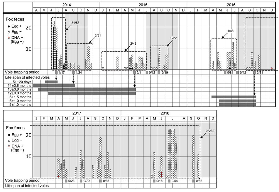
Figure 2. Echinococcus multilocularis tapeworm prevalence in foxes and voles at Hokkaido University campus, Sapporo, Japan, June 2014–October 2018. In the fox feces section, circles in each month show the fecal samples collected at the beginning, middle, and end of the month (88 fecal samples were collected in the middle of July 2018). Black circles indicate fecal samples that were E. multilocularis egg–positive. White circles indicate fecal samples that were E. multilocularis egg–negative. Circled red dots show fecal samples that were egg-negative and positive for E. multilocularis–specific copro-DNA. Fractions indicate the egg-positive rate of fecal samples collected during each period enclosed by a dashed line. Light gray shaded areas indicate the baiting periods. In the vole trapping period section, gray strips show the vole trapping periods. Fractions indicate the infection rate of E. multilocularis in Myodes rufocanus voles in each trapping period. In the life span of infected voles section, dark gray bars show the life span of 7 infected M. rufocanus voles, estimated from the age on the day of trapping (+ SD of the z score). +, positive; –, negative.
1These authors contributed equally to this article.
2Current affiliation: University of Miyazaki, Miyazaki, Japan.
3Current affiliation: Hokkaido University, Sapporo, Japan.