Volume 29, Number 10—October 2023
Dispatch
Angiostrongylus cantonensis Infection in Brown Rats (Rattus norvegicus), Atlanta, Georgia, USA, 2019–2022
Figure
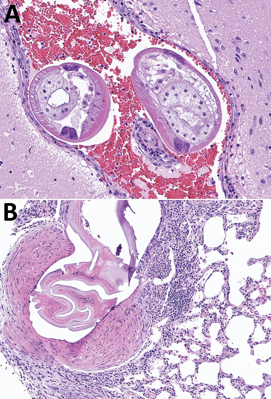
Figure. Brain and lung tissue samples showing Angiostrongylus cantonensis infection in brown rats (Rattus norvegicus), Atlanta, Georgia, USA, 2019–2022. A) Representative tissue section from the brain, stained with hematoxylin and eosin. The meninges and ventricles are multifocally and moderately expanded by abundant hemorrhage. Within the meninges and ventricles, occasional cross sections through nematodes can be seen. Nematodes were ≈250–300 μm in diameter with a thin eosinophilic cuticle, pseudocoelom, polymyarian coelomyarian musculature, lateral chords, and multinucleated intestine. Original magnification ×200 μm. B) Representative tissue section from the lung stained with hematoxylin and eosin. A large pulmonary artery contains fragments of a degenerative nematode characterized by a thin eosinophilic cuticle, pseudocoelomic space, and polymyarian coelomyarian musculature. The subtending arterial wall was sometimes necrotic and variably infiltrated by eosinophils, lymphocytes, and macrophages. The vessel also displays hypertrophy of the tunica media and occasional hypertrophy of the endothelial cells. Original magnification ×100 μm.