Volume 29, Number 11—November 2023
Synopsis
Congenital Mpox Syndrome (Clade I) in Stillborn Fetus after Placental Infection and Intrauterine Transmission, Democratic Republic of the Congo, 2008
David A. Schwartz1 , Placide Mbala-Kingebeni, Kerry Patterson, John W. Huggins, and Phillip R. Pittman1
Figure 3
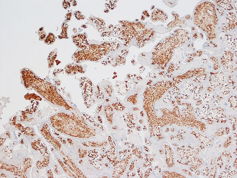
Figure 3. Immunohistochemistry of placenta from a stillborn fetus after placental monkeypox infection and intrauterine transmission, Democratic Republic of the Congo, 2008. Microscopic findings show diffuse and intense positive staining for orthopoxvirus antigen in Hofbauer cells in the chorionic villi. Immunohistochemistry with antibody to vaccinIa virus counterstained with hematoxylin and eosin. Original magnification ×10.
1These senior authors contributed equally to this article.
Page created: September 01, 2023
Page updated: October 23, 2023
Page reviewed: October 23, 2023
The conclusions, findings, and opinions expressed by authors contributing to this journal do not necessarily reflect the official position of the U.S. Department of Health and Human Services, the Public Health Service, the Centers for Disease Control and Prevention, or the authors' affiliated institutions. Use of trade names is for identification only and does not imply endorsement by any of the groups named above.