Skip directly to site content Skip directly to page options Skip directly to A-Z link Skip directly to A-Z link Skip directly to A-Z link
Volume 29, Number 12—December 2023
Research Letter

Tuberculosis in Lemurs and a Fossa at National Zoo, Madagascar, 2022

Marni LaFleurComments to Author , Hoby A. Rasoanaivo, Tojo H. Andrianarivo, Fanantenana Randria Andrianomanana, Stuart McKernan, Mamy Serge Raherison, Rakotoson Andrianantenaina, Michele Miller, Jonah Ratsimbazafy, Simon Grandjean Lapierre, Paulo Ranaivomanana, and Niaina Rakotosamimanana
Author affiliations: University of San Diego, San Diego, California, USA (M. LaFleur); Lemur Love Inc., San Diego (M. LaFleur, H.A. Rasoanaivo); University of Antananarivo, Antananarivo, Madagascar (H. Rasoanaivo, T.H. Andrianarivo); Pasteur Institute of Madagascar, Antananarivo (T.H. Andrianarivo, F.R. Andrianomanana, M.S. Raherison, P. Ranaivomanana, N. Rakotosamimanana); Wildlife One Health, Mtunzini, South Africa (S. McKernan); Programme National de Lutte Contre la Tuberculose, Antananarivo (M.S. Raherison); University of Stellenbosch, Cape Town, South Africa (M. Miller); Centre de Recherche du Centre Hospitalier de l’Université de Montréal, Montréal, Quebec, Canada (S. Grandjean Lapierre); Université de Montréal, Montréal, Canada (S. Grandjean Lapierre)

Main Article

Figure

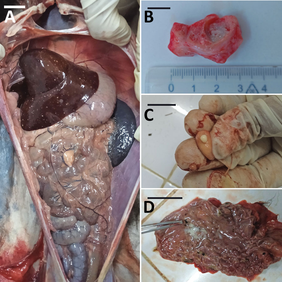
Necropsy images of 2 lemurs (detailed in the Table), both Varecia variegata, from a study of Mycobacterium tuberculosis complex–positive animals at the Botanical and Zoological Park of Tsimbazaza, Madagascar, in 2022. A) Body cavity with nodules and white spots in liver (animal 3). B) Tracheobronchial caseation of lymph nodes (animal 1). C) Tracheobronchial caseation of lymph nodes (animal 1). D) Black spots in stomach mucosa (animal 3). Scale bars indicate 1 cm.

Figure. Necropsy images of 2 lemurs (detailed in the Table), both Varecia variegata, from a study of Mycobacterium tuberculosis complex–positive animals at the Botanical and Zoological Park of Tsimbazaza, Madagascar, in 2022. A) Body cavity with nodules and white spots in liver (animal 3). B) Tracheobronchial caseation of lymph nodes (animal 1). C) Tracheobronchial caseation of lymph nodes (animal 1). D) Black spots in stomach mucosa (animal 3). Scale bars indicate 1 cm.

Main Article

Page created: October 31, 2023
Page updated: November 18, 2023
Page reviewed: November 18, 2023
The conclusions, findings, and opinions expressed by authors contributing to this journal do not necessarily reflect the official position of the U.S. Department of Health and Human Services, the Public Health Service, the Centers for Disease Control and Prevention, or the authors' affiliated institutions. Use of trade names is for identification only and does not imply endorsement by any of the groups named above.
file_external