Volume 29, Number 2—February 2023
Dispatch
Powassan Virus Lineage I in Field-Collected Dermacentor variabilis Ticks, New York, USA
Cite This Article
Citation for Media
Abstract
Powassan virus is a tickborne flavivirus that can cause lethal or debilitating neurologic illness. It is canonically transmitted by Ixodes spp. ticks but might spill over to sympatric Dermacentor species. We detected Powassan virus lineage I from a pool of field-collected D. variabilis ticks in New York, USA.
Powassan virus (POWV) is a neurotropic, tickborne flavivirus first identified as a human pathogen in 1958, when it was isolated from the brain of a patient who had died of encephalitis (1). POWV infection results in febrile illness that can progress to encephalitis, meningitis, and, rarely, meningoencephalitis (2), which is associated with head pain, confusion, paralysis, coma, and death in up to 15% of cases. In addition, >50% of survivors experience long-term neurosequelae, including motor deficiency and cognitive deficits (3).
POWV was initially associated with the woodchuck tick, Ixodes cookei (4), although a second lineage was discovered in deer ticks (I. scapularis) (5). That genotype was termed POWV lineage II, or deer tick virus (DTV). Because of the frequency with which I. scapularis tick bites occur in humans compared to I. cookei tick bites, DTV is likely the most common etiologic source of Powassan encephalitis in the United States. However, the source is difficult to discern because of serologic homology between the virus lineages and the lack of viral genotyping in most clinical settings.
Recently, interest has grown in the vector competency of other sympatric tick species. Dermacentor spp. ticks have been of particular interest because of their common occurrence in POWV- and DTV-endemic areas and because of their tendency to bite humans. POWV has been isolated from D. andersoni ticks in Colorado, USA (6); genetic analysis suggests that strain, called POWV 791A-52, is most likely a form of DTV (7). However, it remains unclear whether the tick in question was infected by spillover from another sylvatic cycle featuring Ixodes ticks or constitutes its own sylvatic system. Neither I. scapularis nor I. cookei ticks are native to Colorado, although several rodent-specific Ixodes species are present (8) that might be involved in such a system.
The competency of D. andersoni ticks for POWV has been confirmed under laboratory conditions when fed from artificially inoculated nonnative species (9). Recent analysis has also indicated that D. variabilis ticks are capable of acquiring and transmitting DTV under laboratory conditions, including maintaining replicating virus transstadially (10). Although that capability has been confirmed in experimentally infected ticks, it remains unclear whether wild populations of D. variabilis ticks can maintain and transmit POWV or DTV under natural circumstances. Considering that D. variabilis ticks are the second most common human-biting species in New York, USA, (11), the ability for the species to transmit POWV in nature represents a critical component of potential human exposure. We detected POWV lineage I from D. variabilis ticks collected in New York in 2021.
As a part of ongoing efforts to track the emergence of POWV in New York, we performed tick surveillance in areas known to contain circulating POWV as identified from a community-engaged tick testing program (11). From 1 area of interest in Dutchess County, New York, we collected 5 female and 3 male D. variabilis ticks, in addition to 68 I. scapularis ticks, in the second half of April 2021. We visually speciated the ticks and assessed them for feeding status. The female D. variabilis ticks were unfed; we pooled, homogenized, and tested them for POWV by quantitative reverse transcription PCR as described (11). In brief, we initially detected POWV with a primer sensitive to both POWV lineage I and DTV. Then, we used a differentiation quantitative reverse transcription PCR to confirm POWV lineage I with a titer of 3.88 log10 FFU/μg RNA. In contrast, none of the I. scapularis ticks collected from the same site tested positive for POWV lineage I and DTV.
We used our highly multiplexed PCR amplicon approach to sequence POWV detected from the tick homogenate (12). We prepared libraries with the Illumina COVIDSeq Test (RUO version; Illumina, https://www.illumina.com), replacing the SARS-CoV-2 primers with POWV (13), and sequenced on the Illumina NovaSeq at the Yale Center for Genome Analysis (New Haven, CT, USA). Consensus genomes were generated at a minimum nucleotide frequency threshold of 0.75 and minimum depth of 10 reads using iVar version 1.3.1 (https://github.com/andersen-lab/ivar).
Figure
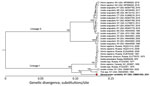
Figure. Maximum-likelihood phylogenetic tree of Powassan virus lineage I and II from Dermacentor variabilisticks collected in New York, USA, and reference sequences. Phylogenetic analysis of the coding sequence (genome...
We reconstructed a maximum-likelihood phylogenetic tree of 29 aligned POWV genomes trimmed to the coding sequence (genome positions 108–10,352) (Figure) using IQ-TREE version 1.6.12 (http://www.iqtree.org) with ultrafast bootstrap approximation (1,000 replicates) (14). Our phylogenetic analysis revealed that the virus (deposited in GenBank under accession no. OM681505) belongs to the POWV lineage I clade and is closely related to a POWV lineage I virus that we sequenced from I. cookei ticks (GenBank accession no. OM681504) from New York in 2020 (Figure). In addition, we used Sanger sequencing of tick ribosomal RNA to confirm that the sample was derived from D. variabilis ticks and not another potential vector species. The resulting sequence (GenBank accession no. ON922563) had 100% homology to D. variabilis large subunit ribosomal RNA (GenBank accession no. L34300.1).
Our results confirm POWV in D. variabilis ticks in southern New York, suggesting that POWV can exist in this tick species, either because of incidental exposure or because of its own sylvatic cycle. In this study, the POWV we identified groups with lineage I; this lineage is normally associated with I. cookei ticks and woodchucks (Marmota monax), instead of with I. scapularis ticks and Peromyscus leucopus mice (15). This link suggests either spillover from that sylvatic cycle, a unique D. variabilis species-dependent sylvatic cycle for POWV lineage I, or a unique subtype of the virus specific to D. variabilis ticks with an unknown sylvatic cycle. Regardless of its source, our data indicate that some D. variabilis ticks in POWV-endemic areas could be capable of acquiring a genotype of POWV similar to lineage I.
POWV is a medically noteworthy flavivirus understood to be primarily transmitted by Ixodes spp. ticks in North America. The sympatric tick species D. variabilis, however, has been recently demonstrated to be a competent vector for POWV under laboratory conditions (10). We report the detection of POWV lineage I in D. variabilis ticks collected from the wild, suggesting that the species might play a direct role in POWV transmission in nature. The ability of POWV to infect humans and the nature of the disease it causes remain unclear, and further research is needed to understand the role of D. variabilis ticks in the ecology of POWV. However, considering that D. variabilis ticks are a primary species of human-biting ticks in New York, this finding demonstrates a new potential source of human exposure to POWV.
Dr. Hart is a postdoctoral researcher at Upstate Medical University in Syracuse, New York, USA. His primary research focus is on the interaction between vector-borne pathogens, arthropod vectors, and vector-host interactions.
Acknowledgment
The study described in this manuscript was funded by Departmental Start-up funds, Upstate Foundation (Fund ID: 23709) and SUNY Empire Innovation Professorship funds to S.T. The funders had no role in study design, data collection and analysis, decision to publish, or preparation of the manuscript.
References
- McLEAN DM, Donohue WL. Powassan virus: isolation of virus from a fatal case of encephalitis. Can Med Assoc J. 1959;80:708–11.PubMedGoogle Scholar
- Gholam BI, Puksa S, Provias JP. Powassan encephalitis: a case report with neuropathology and literature review. CMAJ. 1999;161:1419–22.PubMedGoogle Scholar
- Ebel GD. Update on Powassan virus: emergence of a North American tick-borne flavivirus. Annu Rev Entomol. 2010;55:95–110. DOIPubMedGoogle Scholar
- McLean DM, Best JM, Mahalingam S, Chernesky MA, Wilson WE. Powassan virus: summer infection cycle, 1964. Can Med Assoc J. 1964;91:1360–2.PubMedGoogle Scholar
- Telford SR III, Armstrong PM, Katavolos P, Foppa I, Garcia AS, Wilson ML, et al. A new tick-borne encephalitis-like virus infecting New England deer ticks, Ixodes dammini. Emerg Infect Dis. 1997;3:165–70. DOIPubMedGoogle Scholar
- Thomas LA, Kennedy RC, Eklund CM. Isolation of a virus closely related to Powassan virus from Dermacentor andersoni collected along North Cache la Poudre River, Colo. Proc Soc Exp Biol Med. 1960;104:355–9. DOIPubMedGoogle Scholar
- Robich RM, Cosenza DS, Elias SP, Henderson EF, Lubelczyk CB, Welch M, et al. Prevalence and genetic characterization of deer tick virus (Powassan virus, lineage II) in Ixodes scapularis ticks collected in Maine. Am J Trop Med Hyg. 2019;101:467–71. DOIPubMedGoogle Scholar
- Hutcheson HJ, Mertins JW, Kondratieff BC, White MM. Ticks and tick-borne diseases of Colorado, including new state records for Argas radiatus (Ixodida: Argasidae) and Ixodes brunneus (Ixodida: Ixodidae). J Med Entomol. 2021;58:505–17. DOIPubMedGoogle Scholar
- Chernesky MA. Powassan virus transmission by ixodid ticks infected after feeding on viremic rabbits injected intravenously. Can J Microbiol. 1969;15:521–6. DOIPubMedGoogle Scholar
- Sharma R, Cozens DW, Armstrong PM, Brackney DE. Vector competence of human-biting ticks Ixodes scapularis, Amblyomma americanum and Dermacentor variabilis for Powassan virus. Parasit Vectors. 2021;14:466. DOIPubMedGoogle Scholar
- Hart CE, Bhaskar JR, Reynolds E, Hermance M, Earl M, Mahoney M, et al. Community engaged tick surveillance and tickMAP as a public health tool to track the emergence of ticks and tick-borne diseases in New York. PLOS Glob Public Health. 2022;2:
e0000215 . DOIGoogle Scholar - Grubaugh ND, Gangavarapu K, Quick J, Matteson NL, De Jesus JG, Main BJ, et al. An amplicon-based sequencing framework for accurately measuring intrahost virus diversity using PrimalSeq and iVar. Genome Biol. 2019;20:8. DOIPubMedGoogle Scholar
- Vogels CBF, Brito A, Grubaugh ND. powassan-genomics. 2022 July [cited 2022 Jun 06] https://github.com/grubaughlab/powassan-genomics
- Nguyen LT, Schmidt HA, von Haeseler A, Minh BQ. IQ-TREE: a fast and effective stochastic algorithm for estimating maximum-likelihood phylogenies. Mol Biol Evol. 2015;32:268–74. DOIPubMedGoogle Scholar
- Pesko KN, Torres-Perez F, Hjelle BL, Ebel GD. Molecular epidemiology of Powassan virus in North America. J Gen Virol. 2010;91:2698–705. DOIPubMedGoogle Scholar
Figure
Cite This ArticleOriginal Publication Date: January 13, 2023
1These first authors contributed equally to this article.
Table of Contents – Volume 29, Number 2—February 2023
| EID Search Options |
|---|
|
|
|
|
|
|
Please use the form below to submit correspondence to the authors or contact them at the following address:
Saravanan Thangamani, Upstate Medical University, 505 Irving Ave, Syracuse NY 13210, USA
Top