Volume 29, Number 3—March 2023
Research Letter
Recurrent Cellulitis Revealing Helicobacter cinaedi in Patient on Ibrutinib Therapy, France
Anne-Laure Roupie , Emmanuel Lafont, Sylvie Fraitag, Agnès Ferroni, Hervé Lécuyer, Olivia Boccara, Emilie Bessède, Philippe Lehours, François Lefrère, and Olivier Lortholary
Figure 2
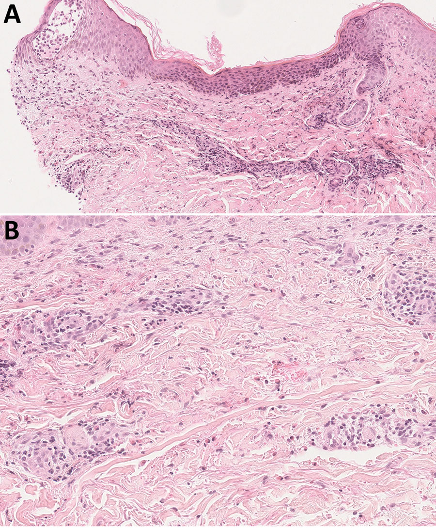
Figure 2. Histologic aspect of a skin lesion revealing Helicobacter cinaedi bacteremia in a man on ibrutinib therapy for chronic lymphocytic leukemia, France. Skin biopsy obtained from erythema on the right side of the abdomen (original magnification ×50 in panel A, ×100 in panel B) show eosinophilic spongiosis leading to spongiotic vesicles and inflammatory perivascular and interstitial infiltrate, mostly located in the superficial and mid dermis and composed of eosinophils and lymphocytes, with no atypical cells. Hematoxylin-eosin-saffron stains.
Page created: January 27, 2023
Page updated: February 20, 2023
Page reviewed: February 20, 2023
The conclusions, findings, and opinions expressed by authors contributing to this journal do not necessarily reflect the official position of the U.S. Department of Health and Human Services, the Public Health Service, the Centers for Disease Control and Prevention, or the authors' affiliated institutions. Use of trade names is for identification only and does not imply endorsement by any of the groups named above.