Volume 29, Number 5—May 2023
Dispatch
No Substantial Histopathologic Changes in Mops condylurus Bats Naturally Infected with Bombali Virus, Kenya
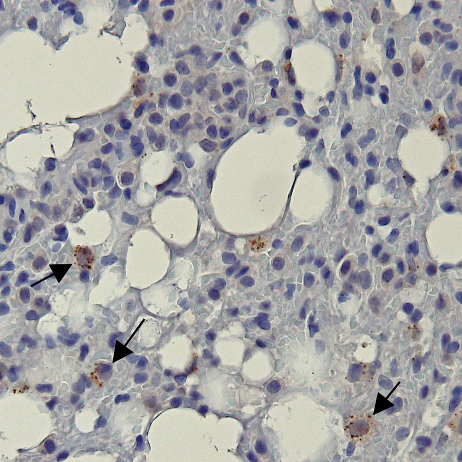
Figure 2

Figure 2. Representative bat lung tissue showing Ebola virus (EBOV) cytoplasmic granules in study of histopathologic changes in Mops condylurus bats naturally infected with Bombali virus, Kenya. We labeled lung tissue sections by using rabbit polyclonal serum against EBOV matrix protein VP40 and detected antigen by using a chromogenic horse radish peroxidase substrate. The sections were then counterstained with hematoxylin. Arrow indicates granular cytoplasmic immunopositivity for EBOV VP40 antigen. Original magnification ×400. VP, viral protein.
Page created: March 06, 2023
Page updated: April 18, 2023
Page reviewed: April 18, 2023
The conclusions, findings, and opinions expressed by authors contributing to this journal do not necessarily reflect the official position of the U.S. Department of Health and Human Services, the Public Health Service, the Centers for Disease Control and Prevention, or the authors' affiliated institutions. Use of trade names is for identification only and does not imply endorsement by any of the groups named above.