Volume 29, Number 7—July 2023
Synopsis
Multicentric Case Series and Literature Review of Coccidioidal Otomastoiditis
Ilan S. Schwartz , Caitlyn Marek, Harleen Sandhu, Ahmed Abdelmonem, Greti Petersen, Emma Dishner, Arash Heidari, and George R. Thompson
Figure 1
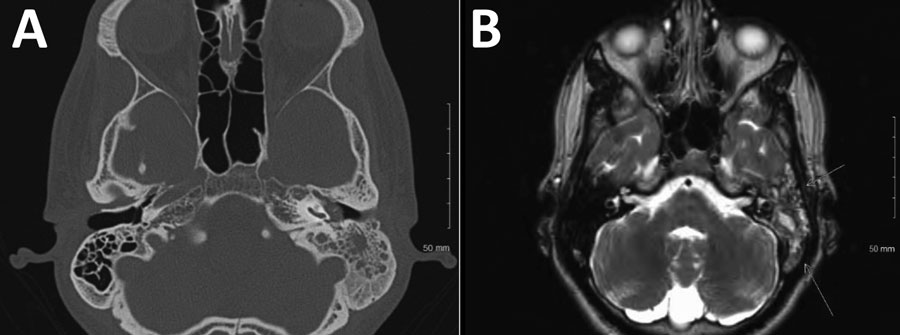
Figure 1. Radiographic findings from 22-year-old Hispanic man from California, USA (case 4), in multicentric case series of coccidioidal otomastoiditis. A) Computed tomography scan of the head, showing opacification of the mastoid. B) Magnetic resonance image of brain, showing mastoiditis.
Page created: May 21, 2023
Page updated: June 21, 2023
Page reviewed: June 21, 2023
The conclusions, findings, and opinions expressed by authors contributing to this journal do not necessarily reflect the official position of the U.S. Department of Health and Human Services, the Public Health Service, the Centers for Disease Control and Prevention, or the authors' affiliated institutions. Use of trade names is for identification only and does not imply endorsement by any of the groups named above.