Volume 29, Number 7—July 2023
Research Letter
Low Susceptibility of Pigs against Experimental Infection with HPAI Virus H5N1 Clade 2.3.4.4b
Cite This Article
Citation for Media
Abstract
We found that nasal and alimentary experimental exposure of pigs to highly pathogenic avian influenza virus H5N1 clade 2.3.4.4b was associated with marginal viral replication, without inducing any clinical manifestation or pathological changes. Only 1 of 8 pigs seroconverted, pointing to high resistance of pigs to clade 2.3.4.4b infection.
Spread of highly pathogenic avian influenza (HPAI) virus H5N1 clade 2.3.4.4b of the goose/Guangdong (gs/GD) lineage, has exacerbated since early 2022 into a panzootic (1). Regional enzootic status in wild bird populations in Europe and North America, with lethal courses of HPAI virus infection in some species, produced large numbers of wild bird carcasses, easy prey for raptors and scavengers. Exposure of terrestrial carnivores and marine mammals resulted in sporadic infections, some of those terminating with fatal encephalitis (2). Frequent spill-over events, rather than consistent mammal-to-mammal transmission, were at the basis of these cases (Figure, panel A). However, recently reported HPAI outbreaks among sea lions along the Pacific coast of South America and an outbreak in a mink farm in Spain (3) may constitute first examples of avian-independent transmission chains and increase public health concerns about zoonotic transmissions of this virus. Still, the total of 11 human cases globally reported for the currently dominating H5N1 2.3.4.4b lineage did not point toward increased zoonotic propensity (4).
Possible adaption of avian influenza virus (AIV) to mammalian livestock hosts and subsequent human exposure is of particular concern. In this respect, the role of pigs as a “mixing vessel” for HPAI viruses is largely unresolved. AIV can potentially be transmitted to pigs, and further reassortment with swine influenza A viruses (swIAVs) may contribute to the emergence of pandemic strains. Rare and subclinical infections of pigs with gs/GD HPAI virus have been confirmed serologically in Vietnam, Thailand, and France (5) and virologically in Indonesia (clades 2.1.1 and 2.1.3), Nigeria (clade 2.3.2.1c), China (clade 2.3.4), and Italy (clade 2.3.4.4.b) (6–8).
Figure
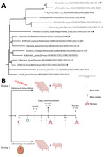
Figure. Phylogeny and experimental design for study of susceptibility of pigs against experimental infection with highly pathogenic avian influenza (HPAI) (H5N1) virus clade 2.3.4.4b. A) Maximum-likelihood phylogenetic tree (RAxML, https://cme.h-its.org/exelixis/web/software/raxml...
For our study, we purchased 4-month-old pigs (6 male, 4 female) from a conventional pig holding in Germany and exposed them nasally or by the alimentary route to high doses of the recent avian-derived HPAI virus H5N1 2.3.4.4b isolate A/chicken/Germany/AI04286/2022 (genotype Ger-10.21-N1.5). The egg-grown isolate was closely related to a mammal case but lacked any mammalian-adaptive mutations (Figure, panel A). We inoculated 2 groups of 4 pigs each intranasally or by feeding 1 infected embryonated chicken egg per animal. One sentinel pig per group was associated at day 1 postinoculation to assess the transmission by direct contact to those inoculated (Figure, panel B).
After exposure, HPAI virus H5N1 detection by real-time RT-PCR was limited to day 2 (intranasal group) and day 4 (oral group) postinoculation, with a range of 10–150 genome copy equivalents per 0.1 mL (Table). An avian-derived, swine-adapted H1avN1 strain intranasally inoculated into naïve pigs at the same dose and by the same device produced 3–4 log10 levels greater nasal excretion in comparison (Table) (9). It cannot be excluded that HPAI virus H5N1 detected in nasal swabs at day 2 postinoculation still represents residual inoculum. Correspondingly, except for 1 alimentary inoculated that showed tracheal viral loads at day 4 postinoculation close to the detection limit, samples from the remaining 3 pigs euthanized at day 4 postinoculation gave no indication of viral replication in respiratory or gut tissues, regardless of the inoculation route. Only in 1 intranasally inoculated animal, euthanized at day 14 postinoculation, was viral RNA detected at low levels in organ samples. In addition to samples from the respiratory and alimentary tracts, minute amounts of RNA were found also in a brain sample of that pig. The virus could not be isolated using chicken hepatoma cells and MDCK-II cells, and histologic and immunohistochemical investigations gave no evidence for inflammatory reactions or presence of viral antigen. Low viral loads in this pig impeded sequence analysis of eventual mutants. Nonetheless, this pig was the only animal that seroconverted at 14 dpi. In agreement with the virologic findings, none of the pigs showed any clinical signs or fever within 14 days of observation.
In conclusion, only 1 of 8 pigs inoculated intranasally with HPAI virus H5N1 underwent transient, low-level infection that resulted in the presence of viral RNA in several tissue specimens and seroconversion at 14 dpi. In naturally infected wild mammals, this virus was prominently detected in the brain (2). Given the detection of viral RNA in the brain of 1 intranasally inoculated pig, it cannot be excluded that longer observation might have revealed continuing viral replication in the brain of this animal. Sialic acid α2,3-gal receptors dominate on porcine brain cells, which might have fostered replication of α2,3-adapted viruses, such as HPAI virus H5N1 (10).
Overall, we conclude that pigs are unlikely vehicles in transmitting this genotype of HPAI virus H5N1 clade 2.3.4.4b among pigs and across interfaces. However, considering the ongoing massive panzootic of this virus, a plethora of new genotypes of the circulating strain is emerging, with possibly higher permissiveness for pigs. Therefore, swine populations need to be part of HPAI virus surveillance programs, and periodic reassessment of prepandemic propensity of circulating HPAI virus H5N1 genotypes in the swine model is required.
Dr. Graaf is a veterinary virologist at the International (World Organisation for Animal Health/Food and Agriculture Organization of the United Nations) and National Reference Laboratory for Avian Influenza at the Institute of Diagnostic Virology at the Friedrich-Loeffler-Institute. Her research interests focus on avian and swine influenza A virus surveillance in Germany.
Acknowledgments
We acknowledge the originating and submitting laboratories that provided sequences available in the GISAID EpiFlu database (http://www.gisaid.org). We are grateful to animal keepers Doreen Fiedler, Frank Klipp, Christian Lipinski, and Steffen Kiepert.
Animal experiments were approved by the State Office for Agriculture, Food Safety and Fishery, in the Federal State of Mecklenburg-Western Pomerania, Germany (LALFF M-V 7221.3-2-010/18). All animals were kept under BioSafety Level 3 conditions in the corresponding animal facilities at Friedrich-Loeffler-Institute, Greifswald–Insel Riems, Germany.
References
- Alkie TN, Lopes S, Hisanaga T, Xu W, Suderman M, Koziuk J, et al. A threat from both sides: Multiple introductions of genetically distinct H5 HPAI viruses into Canada via both East Asia-Australasia/Pacific and Atlantic flyways. Virus Evol. 2022;8:veac077.
- Bordes L, Vreman S, Heutink R, Roose M, Venema S, Pritz-Verschuren SBE, et al. Highly pathogenic avian influenza H5N1 virus infections in wild red foxes (Vulpes vulpes) show neurotropism and adaptive virus mutations. Microbiol Spectr. 2023;11:
e0286722 . DOIPubMedGoogle Scholar - Agüero M, Monne I, Sánchez A, Zecchin B, Fusaro A, Ruano MJ, et al. Highly pathogenic avian influenza A(H5N1) virus infection in farmed minks, Spain, October 2022. Euro Surveill. 2023;28:5–10. DOIPubMedGoogle Scholar
- Centers for Disease Control and Prevention. Ask the expert: highly pathogenic avian influenza A (H5N1) viruses [cited 2023 Apr 14]. https://www.cdc.gov/flu/avianflu/spotlights/2022-2023/avian-flu-highly-pathogenic.htm
- Choi YK, Nguyen TD, Ozaki H, Webby RJ, Puthavathana P, Buranathal C, et al. Studies of H5N1 influenza virus infection of pigs by using viruses isolated in Vietnam and Thailand in 2004. J Virol. 2005;79:10821–5. DOIPubMedGoogle Scholar
- He L, Zhao G, Zhong L, Liu Q, Duan Z, Gu M, et al. Isolation and characterization of two H5N1 influenza viruses from swine in Jiangsu Province of China. Arch Virol. 2013;158:2531–41. DOIPubMedGoogle Scholar
- Meseko C, Globig A, Ijomanta J, Joannis T, Nwosuh C, Shamaki D, et al. Evidence of exposure of domestic pigs to Highly Pathogenic Avian Influenza H5N1 in Nigeria. Sci Rep. 2018;8:5900. DOIPubMedGoogle Scholar
- Nidom CA, Takano R, Yamada S, Sakai-Tagawa Y, Daulay S, Aswadi D, et al. Influenza A (H5N1) viruses from pigs, Indonesia. Emerg Infect Dis. 2010;16:1515–23. DOIPubMedGoogle Scholar
- Graaf A, Petric PP, Sehl-Ewert J, Henritzi D, Breithaupt A, King J, et al. Cold-passaged isolates and bat-swine influenza a chimeric viruses as modified live-attenuated vaccines against influenza a viruses in pigs. Vaccine. 2022;40:6255–70. DOIPubMedGoogle Scholar
- Nelli RK, Kuchipudi SV, White GA, Perez BB, Dunham SP, Chang KC. Comparative distribution of human and avian type sialic acid influenza receptors in the pig. BMC Vet Res. 2010;6:4. DOIPubMedGoogle Scholar
Figure
Table
Cite This ArticleOriginal Publication Date: June 14, 2023
Table of Contents – Volume 29, Number 7—July 2023
| EID Search Options |
|---|
|
|
|
|
|
|
Please use the form below to submit correspondence to the authors or contact them at the following address:
Annika Graaf, Friedrich-Loeffler-Institute, Suedufer 1017493, Greifswald–Insel Riems, Germany
Top