Clinical Characteristics of Corynebacterium ulcerans Infection, Japan
Akihiko Yamamoto

, Toru Hifumi, Manabu Ato, Masaaki Iwaki, Mitsutoshi Senoh, Akio Hatanaka, Shinichi Nureki, Yoshihiro Noguchi, Tomoko Hirose, Yukihiro Yoshimura, Takaaki Urakawa, Shiro Hori, Hiroto Nakada, Tomomasa Terada, Tomoko Ishifuji, Hisayo Matsuyama, Takahiro Kinebuchi, Atsuhito Fukushima, Koji Wake, Ken Otsuji, Takeru Endo, Hirokazu Toyoshima, Ikkoh Yasuda, Takeshi Tanaka, Naoki Takahashi, Kensaku Okada, Toshimasa Hayashi, Taizo Kusano, Minami Koriyama, Norio Otani, and Motohide Takahashi
Author affiliations: National Institute of Infectious Diseases, Tokyo, Japan (A. Yamamoto, M. Ato, M. Iwaki, M. Senoh); St. Luke’s International Hospital, Tokyo (T. Hifumi, N. Otani); Ageo Central Genral Hospital, Saitama, Japan (A. Hatanaka); Oita University Faculty of Medicine, Oita, Japan (S. Nureki); International University of Health and Welfare School of Medicine, Chiba, Japan (Y. Noguchi); Japanese Red Cross Otsu Hospital, Shiga, Japan (T. Hirose); Yokohama Municipal Citizen’s Hospital, Kanagawa, Japan (Y. Yoshimura); Tsuruoka Municipal Shonai Hospital, Yamagata, Japan (T. Urakawa); Japan Community Healthcare Organization Ritsurin Hospital, Kagawa, Japan (S. Hori); Holon Toriizaka Clinic, Tokyo (H. Nakada); Tokushima Prefectural Central Hospital, Tokushima, Japan (T. Terada); Itabashi Medical System Tokyo–Katsushika General Hospital, Katsushika, Tokyo (T. Ishifuji); Kawakita General Hospital, Suginami, Tokyo (H. Matsuyama); Furano Hospital, Hokkaido, Japan (T. Kinebuchi); Dokkyo Medical University, Tochigi, Japan (A. Fukushima, K. Wake); Hospital of University of Occupational and Environmental Health, Fukuoka, Japan (K. Otsuji, T. Endo); Japanese Red Cross Ise Hospital, Mie, Japan (H. Toyoshima); Fukushima Medical University, Fukushima, Japan (I. Yasuda); Nagasaki University Hospital, Nagasaki, Japan (T. Tanaka); Kimitsu Chuo Hospital, Chiba (N. Tanaka); Tottori University Hospital, Tottori, Japan (K. Okada); Maebashi Red Cross Hospital, Gunma, Japan (T. Hayashi); Chiba Children’s Hospital, Chiba (T. Kusano); Chiba Rousai Hospital, Chiba (M. Koriyama); Kumamoto Health Science University, Kumamoto, Japan (M. Takahashi)
Main Article
Figure 4

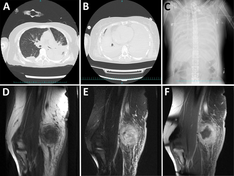
Figure 4. Computed tomography, radiograph, and magnetic resonance imaging results for patients with Corynebacterium ulcerans infection, Japan, 2001–2020. A–C) Chest computed tomography images and radiograph of patients with severe respiratory symptoms. Atelectasis noted at admission (A, top) and after exacerbation of symptoms (B, bottom) (case no. 29, from Dr. Hayashi, Maebashi Red Cross Hospital, Gunma, Japan). Spread of atelectasis noted on chest radiograph taken at the time of exacerbation of symptoms (C) (case no. 29, also from Dr. Hayashi, Maebashi Red Cross Hospital). D–F) Magnetic resonance imaging of an elbow abscess. Magnetic resonance imaging T1-weighted images show equal brightness to the muscles (D), fat-suppressed T2-weighted image by short-tau inversion recovery method show unevenly high brightness (E), and contrast-enhanced T1-weighted images show a mass whose margins are contrast-enhanced (F) (case no. 10, from Dr. Urakawa, Tsuruoka Municipal Shonai Hospital, Yamagata, Japan).
Main Article
Page created: May 31, 2023
Page updated: July 20, 2023
Page reviewed: July 20, 2023
The conclusions, findings, and opinions expressed by authors contributing to this journal do not necessarily reflect the official position of the U.S. Department of Health and Human Services, the Public Health Service, the Centers for Disease Control and Prevention, or the authors' affiliated institutions. Use of trade names is for identification only and does not imply endorsement by any of the groups named above.
