Volume 30, Number 10—October 2024
Research Letter
Establishment of Amblyomma maculatum Ticks and Rickettsia parkeri in the Northeastern United States
Goudarz Molaei , Noelle Khalil, Carmen J. Ramos, and Christopher D. Paddock
Figure 2
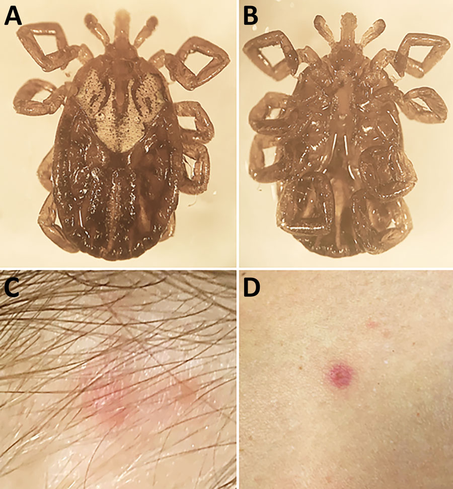
Figure 2. Biting Amblyomma maculatum tick removed from a woman in Connecticut, USA, and signs of Rickettsia parkeri rickettsiosis. A, B) Dorsal (A) and ventral (B) images of the tick. C, D) A small, erythematous, crusted lesion with a smaller satellite papule that developed at the bite site (C) and 1 of several small erythematous macules that developed on her arm and legs (D).
Page created: August 08, 2024
Page updated: September 24, 2024
Page reviewed: September 24, 2024
The conclusions, findings, and opinions expressed by authors contributing to this journal do not necessarily reflect the official position of the U.S. Department of Health and Human Services, the Public Health Service, the Centers for Disease Control and Prevention, or the authors' affiliated institutions. Use of trade names is for identification only and does not imply endorsement by any of the groups named above.