Volume 30, Number 12—December 2024
Research
Highly Pathogenic Avian Influenza A(H5N1) Virus Infection in Cats, South Korea, 2023
Figure 1

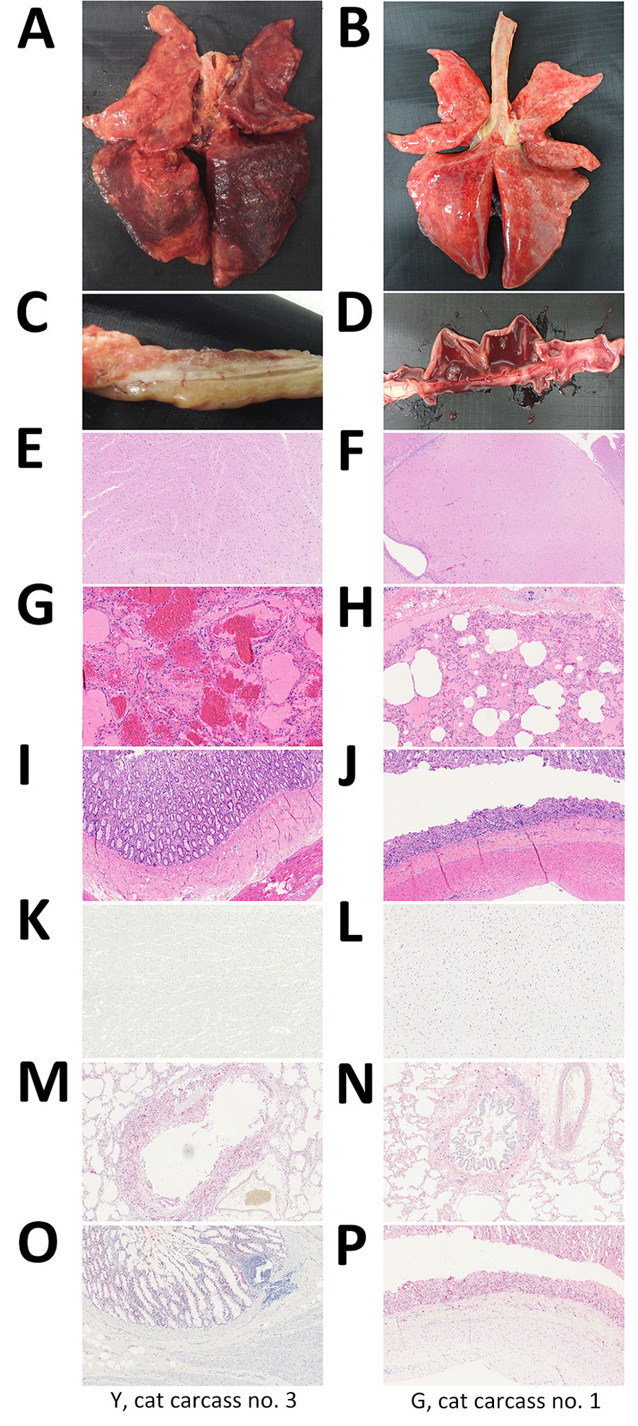
Figure 1. Gross, microscopic, and immunohistochemistry (IHC) findings in cats infected with highly pathogenic avian influenza A(H5N1) virus, South Korea, 2023. Findings are shown for cat carcasses from shelter 1 (Y cat carcass no. 3) and shelter 2 (G cat carcass no. 1). A–D) Gross findings: A) severe congestion and edema in the lungs; B) congestion and edema in the lungs; C) lack of lesions in the small intestine; D) bloody diarrhea in the small intestine (D). E–J) Hemotoxylin and eosin staining: E) brain showing no lesions; F) multifocal gliosis in the brain; G) interstitial pneumonia with focally extensive vascular thrombosis; H) interstitial pneumonia characterized by invasion of the alveolar lumina by mixed neutrophils and macrophages; I) intestine showing no lesions; J) necrotic enteritis with denuded villi. K–P) Immunohistochemical staining: K) brain showing no influenza virus antigens; L) influenza virus antigens in the neurons; M) influenza virus antigens in alveolar macrophages and bronchial epithelial cells; N) influenza virus antigens in alveolar macrophages and bronchial epithelial cells; O) influenza virus antigens in the small intestine; P) influenza virus antigens in the crypt epithelium and blood vessels in the submucosa. Original magnification ×100, except panel F, in which original magnification was ×10.
1These authors contributed equally to this article.