Volume 30, Number 12—December 2024
Dispatch
Experimental Infection of Reindeer with Jamestown Canyon Virus
Figure 3
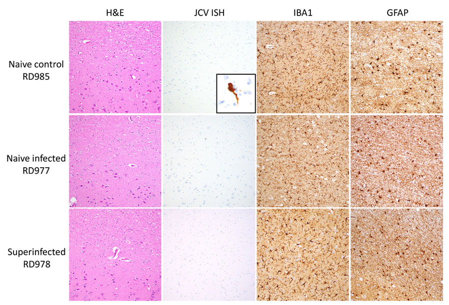
Figure 3. Histopathology of samples from reindeer experimentally infected with JCV. Representative brain sections from naive control (RD985), naive infected (RD977), and superinfected (RD978) animals shows H&E-stained sections of cerebral cortex and subcortical white matter with minimal diagnostic abnormalities, negative JCV RNA ISH staining, scattered IBA1-positive microglia/macrophages, and mild-to-moderate astrocytosis highlighted by GFAP immunohistochemistry. JCV ISH naive control inset panel demonstrates positive cytoplasmic staining in a cortical neuron from a positive control case of fatal JCV encephalitis in a human. All images taken with 20× objective with exception of the JCV ISH inset, taken with 60× objective. GFAP, glial fibrillary acidic protein; H&E, hematoxylin and eosin; IBA, allograft inflammatory factor 1; ISH, in situ hybridization; JCV, Jamestown Canyon virus.