Skip directly to site content Skip directly to page options Skip directly to A-Z link Skip directly to A-Z link Skip directly to A-Z link
Volume 30, Number 2—February 2024
Dispatch

Borrelia turicatae from Ticks in Peridomestic Setting, Camayeca, Mexico

Edwin Vázquez-Guerrero, Alexander R. Kneubehl, Patricio Pellegrini-Hernández, José Luis González-Quiroz, María Lilia Domínguez-López, Aparna Krishnavajhala, Paulina Estrada-de los Santos, J. Antonio IbarraComments to Author , and Job E. Lopez
Author affiliations: Instituto Politécnico Nacional, Mexico City, Mexico (E. Vázquez-Guerrero, J.L. González-Quiroz, M.L. Domínguez-López, P. Estrada-de los Santos, J.A. Ibarra); Baylor College of Medicine, Houston, Texas, USA (A.R. Kneubehl, A. Krishnavajhala, J.E. Lopez); Wildlife Conservation Management Unit (Macochín), El Fuerte, Sinaloa, Mexico (P. Pellegrini-Hernández).

Main Article

Figure 2

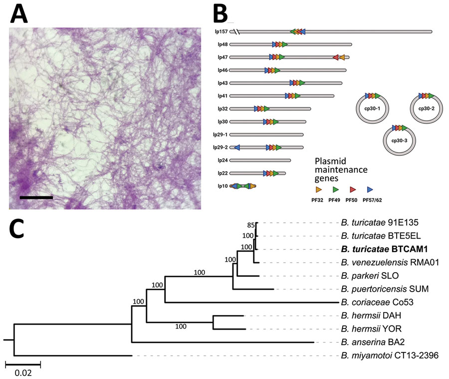
Isolation and genetic characterization of Borrelia turicatae from ticks collected in the village of Camayeca, Mexico. A) Spirochetes were isolated from murine blood in culture medium. Scale bar indicates 20 µm. B) Genome sequencing and assembly generated the plasmid repertoire of the bacteria. Plasmids were designated as lp or cp and by their respective size to the nearest kilobase. PF partitioning genes are shown in each plasmid as orange, green, red, and blue triangles. C) Maximum-likelihood species tree performed in a phylogenomic analysis of the spirochete sample we extracted, designated CAM-1 (boldface), grouped the spirochete with B. turicatae. The tree was generated with an edge-linked proportional partition model with 1,000 ultra-fast bootstraps. Scale bar indicates 0.02 substitutions per site. cp, circular plasmid; lp, linear plasmid; PF, plasmid family.

Figure 2. Isolation and genetic characterization of Borrelia turicatae from ticks collected in the village of Camayeca, Mexico. A) Spirochetes were isolated from murine blood in culture medium. Scale bar indicates 20 µm. B) Genome sequencing and assembly generated the plasmid repertoire of the bacteria. Plasmids were designated as lp or cp and by their respective size to the nearest kilobase. PF partitioning genes are shown in each plasmid as orange, green, red, and blue triangles. C) Maximum-likelihood species tree performed in a phylogenomic analysis of the spirochete sample we extracted, designated CAM-1 (boldface), grouped the spirochete with B. turicatae. The tree was generated with an edge-linked proportional partition model with 1,000 ultra-fast bootstraps. Scale bar indicates 0.02 substitutions per site. cp, circular plasmid; lp, linear plasmid; PF, plasmid family.

Main Article

Page created: January 05, 2024
Page updated: January 24, 2024
Page reviewed: January 24, 2024
The conclusions, findings, and opinions expressed by authors contributing to this journal do not necessarily reflect the official position of the U.S. Department of Health and Human Services, the Public Health Service, the Centers for Disease Control and Prevention, or the authors' affiliated institutions. Use of trade names is for identification only and does not imply endorsement by any of the groups named above.
file_external